

Dendrites
Nucleolus
Axon
Soma (cell body)
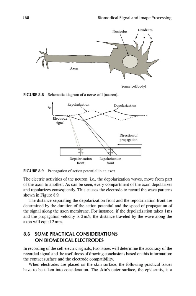
FIGURE 8.8 Schematic diagram of a nerve cell (neuron).
Repolarization
Depolarization
ε m
Electrode
signal
Direction of
propagation
+ –
+ –
– +
+
–
Depolarization
Repolarization
front
front
168
Biomedical Signal and Image Processing
FIGURE 8.9 Propagation of action potential in an axon.
The electric activities of the neuron, i.e., the depolarization waves, move from part
of the axon to another. As can be seen, every compartment of the axon depolarizes
and repolarizes consequently. This causes the electrode to record the wave patterns
shown in Figure 8.9.
The distance separating the depolarization front and the repolarization front are
determined by the duration of the action potential and the speed of propagation of
the signal along the axon membrane. For instance, if the depolarization takes 1 ms
and the propagation velocity is 2 m/s, the distance traveled by the wave along the
axon will equal 2 mm.
8.6
SOME PRACTICAL CONSIDERATIONS
ON BIOMEDICAL ELECTRODES
In recording of the cell electric signals, two issues will determine the accuracy of the
recorded signal and the usefulness of drawing conclusions based on this information:
the contact surface and the electrode compatibility.
When electrodes are placed on the skin surface, the following practical issues
have to be taken into consideration. The skin’s outer surface, the epidermis, is a
Nucleolus
Axon
Soma (cell body)
FIGURE 8.8 Schematic diagram of a nerve cell (neuron).
Repolarization
Depolarization
ε m
Electrode
signal
Direction of
propagation
+ –
+ –
– +
+
–
Depolarization
Repolarization
front
front
168
Biomedical Signal and Image Processing
FIGURE 8.9 Propagation of action potential in an axon.
The electric activities of the neuron, i.e., the depolarization waves, move from part
of the axon to another. As can be seen, every compartment of the axon depolarizes
and repolarizes consequently. This causes the electrode to record the wave patterns
shown in Figure 8.9.
The distance separating the depolarization front and the repolarization front are
determined by the duration of the action potential and the speed of propagation of
the signal along the axon membrane. For instance, if the depolarization takes 1 ms
and the propagation velocity is 2 m/s, the distance traveled by the wave along the
axon will equal 2 mm.
8.6
SOME PRACTICAL CONSIDERATIONS
ON BIOMEDICAL ELECTRODES
In recording of the cell electric signals, two issues will determine the accuracy of the
recorded signal and the usefulness of drawing conclusions based on this information:
the contact surface and the electrode compatibility.
When electrodes are placed on the skin surface, the following practical issues
have to be taken into consideration. The skin’s outer surface, the epidermis, is a