

167
Electric Activities of the Cell
(a)
(b)
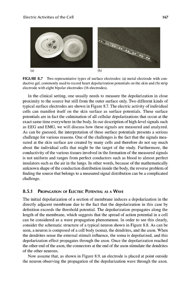
FIGURE 8.7 Two representative types of surface electrodes: (a) metal electrode with conductive gel, commonly used to record heart depolarization potentials on the skin and (b) strip
electrode with eight bipolar electrodes (16 electrodes).
In the clinical setting, one usually needs to measure the depolarization in close
proximity to the source but still from the outer surface only. Two different kinds of
typical surface electrodes are shown in Figure 8.7. The electric activity of individual
cells can manifest itself on the skin surface as surface potentials. These surface
potentials are in fact the culmination of all cellular depolarizations that occur at the
exact same time everywhere in the body. In our description of high-level signals such
as EEG and EMG, we will discuss how these signals are measured and analyzed.
As can be guessed, the interpretation of these surface potentials presents a serious
challenge for various reasons. One of the challenges is the fact that the signals measured at the skin surface are created by many cells and therefore do not say much
about the individual cells that might be the target of the study. Furthermore, the
conductivity of the various tissues involved in the formation of the measured signals
is not uniform and ranges from perfect conductors such as blood to almost perfect
insulators such as the air in the lungs. In other words, because of the mathematically
unknown shape of the conduction distribution inside the body, the reverse problem of
finding the source that belongs to a measured signal distribution can be a complicated
challenge.
8.5.1 PROPAGATION OF ELECTRIC POTENTIAL AS A WAVE
The initial depolarization of a section of membrane induces a depolarization in the
directly adjacent membrane due to the fact that the depolarization in this case by
definition exceeds the threshold potential. The depolarization propagates along the
length of the membrane, which suggests that the spread of action potential in a cell
can be considered as a wave propagation phenomenon. In order to see this clearly,
consider the schematic structure of a typical neuron shown in Figure 8.8. As can be
seen, a neuron is composed of a cell body (soma), the dendrites, and the axon. When
the dendrites sense the external stimuli influence, the soma is depolarized, and this
depolarization effect propagates through the axon. Once the depolarization reached
the other end of the axon, the connectors at the end of the axon stimulate the dendrites
of the other neurons.
Now assume that, as shown in Figure 8.9, an electrode is placed at point outside
the neuron observing the propagation of the depolarization wave through the axon.
Electric Activities of the Cell
(a)
(b)
FIGURE 8.7 Two representative types of surface electrodes: (a) metal electrode with conductive gel, commonly used to record heart depolarization potentials on the skin and (b) strip
electrode with eight bipolar electrodes (16 electrodes).
In the clinical setting, one usually needs to measure the depolarization in close
proximity to the source but still from the outer surface only. Two different kinds of
typical surface electrodes are shown in Figure 8.7. The electric activity of individual
cells can manifest itself on the skin surface as surface potentials. These surface
potentials are in fact the culmination of all cellular depolarizations that occur at the
exact same time everywhere in the body. In our description of high-level signals such
as EEG and EMG, we will discuss how these signals are measured and analyzed.
As can be guessed, the interpretation of these surface potentials presents a serious
challenge for various reasons. One of the challenges is the fact that the signals measured at the skin surface are created by many cells and therefore do not say much
about the individual cells that might be the target of the study. Furthermore, the
conductivity of the various tissues involved in the formation of the measured signals
is not uniform and ranges from perfect conductors such as blood to almost perfect
insulators such as the air in the lungs. In other words, because of the mathematically
unknown shape of the conduction distribution inside the body, the reverse problem of
finding the source that belongs to a measured signal distribution can be a complicated
challenge.
8.5.1 PROPAGATION OF ELECTRIC POTENTIAL AS A WAVE
The initial depolarization of a section of membrane induces a depolarization in the
directly adjacent membrane due to the fact that the depolarization in this case by
definition exceeds the threshold potential. The depolarization propagates along the
length of the membrane, which suggests that the spread of action potential in a cell
can be considered as a wave propagation phenomenon. In order to see this clearly,
consider the schematic structure of a typical neuron shown in Figure 8.8. As can be
seen, a neuron is composed of a cell body (soma), the dendrites, and the axon. When
the dendrites sense the external stimuli influence, the soma is depolarized, and this
depolarization effect propagates through the axon. Once the depolarization reached
the other end of the axon, the connectors at the end of the axon stimulate the dendrites
of the other neurons.
Now assume that, as shown in Figure 8.9, an electrode is placed at point outside
the neuron observing the propagation of the depolarization wave through the axon.