

3.5 Binding of Cysteine with Dopaquinone
75
bond length), indicating a less ordinary covalent bonding unlike the other cases; for
instance, the C5-bound structure (a) shows 1.91 Å of C5–S bond length. The unusual
covalent nature of the C3–C4-bound structure (d) also manifests in a relatively small
geometrical alteration upon binding unlike the other cases (a, b, c, and e) exhibiting
a sp
2 -to-sp
3 (planar-to-pyramidal) structural change during the reaction.
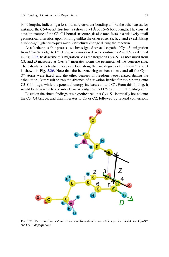
As a further possible process, we investigated a reaction path of Cys–S
− migration
from C3–C4 bridge to C5. Then, we considered two coordinates Z and D, as defined
in Fig. 3.25, to describe this migration. Z is the height of Cys–S
− as measured from
C3, and D increases as Cys–S
− migrates along the perimeter of the benzene ring.
The calculated potential energy surface along the two degrees of freedom Z and D
is shown in Fig. 3.26. Note that the benzene ring carbon atoms, and all the Cys–
S
− atoms were fixed, and the other degrees of freedom were relaxed during the
calculation. Our result shows the absence of activation barrier for the binding onto
C3–C4 bridge, while the potential energy increases around C5. From this finding, it
would be advisable to consider C3–C4 bridge but not C5 as the initial binding site.
Based on the above findings, we hypothesized that Cys–S
− is initially bound onto
the C3–C4 bridge, and then migrates to C5 or C2, followed by several conversions
Fig. 3.25 Two coordinates Z and D for bond formation between S in cysteine thiolate ion Cys-S −
and C5 in dopaquinone
75
bond length), indicating a less ordinary covalent bonding unlike the other cases; for
instance, the C5-bound structure (a) shows 1.91 Å of C5–S bond length. The unusual
covalent nature of the C3–C4-bound structure (d) also manifests in a relatively small
geometrical alteration upon binding unlike the other cases (a, b, c, and e) exhibiting
a sp
2 -to-sp
3 (planar-to-pyramidal) structural change during the reaction.
As a further possible process, we investigated a reaction path of Cys–S
− migration
from C3–C4 bridge to C5. Then, we considered two coordinates Z and D, as defined
in Fig. 3.25, to describe this migration. Z is the height of Cys–S
− as measured from
C3, and D increases as Cys–S
− migrates along the perimeter of the benzene ring.
The calculated potential energy surface along the two degrees of freedom Z and D
is shown in Fig. 3.26. Note that the benzene ring carbon atoms, and all the Cys–
S
− atoms were fixed, and the other degrees of freedom were relaxed during the
calculation. Our result shows the absence of activation barrier for the binding onto
C3–C4 bridge, while the potential energy increases around C5. From this finding, it
would be advisable to consider C3–C4 bridge but not C5 as the initial binding site.
Based on the above findings, we hypothesized that Cys–S
− is initially bound onto
the C3–C4 bridge, and then migrates to C5 or C2, followed by several conversions
Fig. 3.25 Two coordinates Z and D for bond formation between S in cysteine thiolate ion Cys-S −
and C5 in dopaquinone