

3.4 Cyclization of Dopamine Quinone Analogs
73
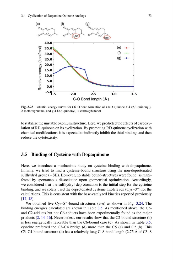
Fig. 3.23 Potential energy curves for C6–O bond formation of e RD-quinone, f 4-(2,3-quinonyl)2-methoxybutane, and g 4-(2,3-quinonyl)-2-carboxybutanol
to stabilize the unstable oxonium structure. Here, we predicted the effects of carboxylation of RD-quinone on its cyclization. By promoting RD-quinone cyclization with
chemical modifications, it is expected to indirectly inhibit the thiol binding, and then
reduce the cytotoxicity.
3.5 Binding of Cysteine with Dopaquinone
Here, we introduce a mechanistic study on cysteine binding with dopaquinone.
Initially, we tried to find a cysteine-bound structure using the non-deprotonated
sulfhydryl group (−SH). However, no stable bound-structures were found, as manifested by spontaneous dissociation upon geometrical optimization. Accordingly,
we considered that the sulfhydryl deprotonation is the initial step for the cysteine
binding, and we solely used the deprotonated cysteine thiolate ion (Cys–S
− ) for the
calculations. This is consistent with the base-catalyzed kinetics reported previously
[17, 18].
We obtained five Cys–S
− -bound structures (a–e) as shown in Fig. 3.24. The
binding energies calculated are shown in Table 3.5. As mentioned above, the C5and C2-adducts but not C6-adducts have been experimentally found as the major
products [2, 14–16]. Nevertheless, our results show that the C2-bound structure (b)
is less energetically favorable than the C6-bound case (c). As shown in Table 3.5,
cysteine preferred the C3–C4 bridge (d) more than the C5 (a) and C2 (b). This
C3–C4-bound structure (d) has a relatively long C–S bond length (2.75 Å of C3–S
73
Fig. 3.23 Potential energy curves for C6–O bond formation of e RD-quinone, f 4-(2,3-quinonyl)2-methoxybutane, and g 4-(2,3-quinonyl)-2-carboxybutanol
to stabilize the unstable oxonium structure. Here, we predicted the effects of carboxylation of RD-quinone on its cyclization. By promoting RD-quinone cyclization with
chemical modifications, it is expected to indirectly inhibit the thiol binding, and then
reduce the cytotoxicity.
3.5 Binding of Cysteine with Dopaquinone
Here, we introduce a mechanistic study on cysteine binding with dopaquinone.
Initially, we tried to find a cysteine-bound structure using the non-deprotonated
sulfhydryl group (−SH). However, no stable bound-structures were found, as manifested by spontaneous dissociation upon geometrical optimization. Accordingly,
we considered that the sulfhydryl deprotonation is the initial step for the cysteine
binding, and we solely used the deprotonated cysteine thiolate ion (Cys–S
− ) for the
calculations. This is consistent with the base-catalyzed kinetics reported previously
[17, 18].
We obtained five Cys–S
− -bound structures (a–e) as shown in Fig. 3.24. The
binding energies calculated are shown in Table 3.5. As mentioned above, the C5and C2-adducts but not C6-adducts have been experimentally found as the major
products [2, 14–16]. Nevertheless, our results show that the C2-bound structure (b)
is less energetically favorable than the C6-bound case (c). As shown in Table 3.5,
cysteine preferred the C3–C4 bridge (d) more than the C5 (a) and C2 (b). This
C3–C4-bound structure (d) has a relatively long C–S bond length (2.75 Å of C3–S