

64
3 Dopaquinone Conversion and Related Reactions
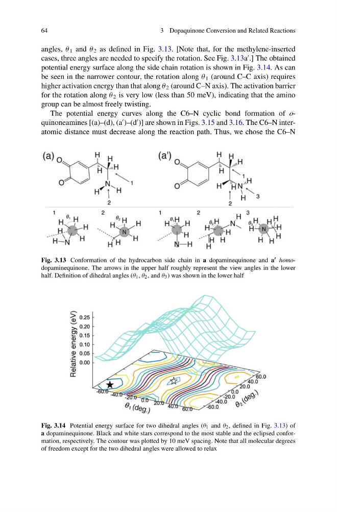
angles, θ 1 and θ 2 as defined in Fig. 3.13. [Note that, for the methylene-inserted
cases, three angles are needed to specify the rotation. See Fig. 3.13a
.] The obtained
potential energy surface along the side chain rotation is shown in Fig. 3.14. As can
be seen in the narrower contour, the rotation along θ 1 (around C–C axis) requires
higher activation energy than that along θ 2 (around C–N axis). The activation barrier
for the rotation along θ 2 is very low (less than 50 meV), indicating that the amino
group can be almost freely twisting.
The potential energy curves along the C6–N cyclic bond formation of oquinoneamines [(a)–(d), (a
)–(d
)] are shown in Figs. 3.15 and 3.16. The C6–N interatomic distance must decrease along the reaction path. Thus, we chose the C6–N
Fig. 3.13 Conformation of the hydrocarbon side chain in a dopaminequinone and a homodopaminequinone. The arrows in the upper half roughly represent the view angles in the lower
half. Definition of dihedral angles (θ 1 , θ 2 , and θ 3 ) was shown in the lower half
Fig. 3.14 Potential energy surface for two dihedral angles (θ 1 and θ 2 , defined in Fig. 3.13) of
a dopaminequinone. Black and white stars correspond to the most stable and the eclipsed conformation, respectively. The contour was plotted by 10 meV spacing. Note that all molecular degrees
of freedom except for the two dihedral angles were allowed to relax
3 Dopaquinone Conversion and Related Reactions
angles, θ 1 and θ 2 as defined in Fig. 3.13. [Note that, for the methylene-inserted
cases, three angles are needed to specify the rotation. See Fig. 3.13a
.] The obtained
potential energy surface along the side chain rotation is shown in Fig. 3.14. As can
be seen in the narrower contour, the rotation along θ 1 (around C–C axis) requires
higher activation energy than that along θ 2 (around C–N axis). The activation barrier
for the rotation along θ 2 is very low (less than 50 meV), indicating that the amino
group can be almost freely twisting.
The potential energy curves along the C6–N cyclic bond formation of oquinoneamines [(a)–(d), (a
)–(d
)] are shown in Figs. 3.15 and 3.16. The C6–N interatomic distance must decrease along the reaction path. Thus, we chose the C6–N
Fig. 3.13 Conformation of the hydrocarbon side chain in a dopaminequinone and a homodopaminequinone. The arrows in the upper half roughly represent the view angles in the lower
half. Definition of dihedral angles (θ 1 , θ 2 , and θ 3 ) was shown in the lower half
Fig. 3.14 Potential energy surface for two dihedral angles (θ 1 and θ 2 , defined in Fig. 3.13) of
a dopaminequinone. Black and white stars correspond to the most stable and the eclipsed conformation, respectively. The contour was plotted by 10 meV spacing. Note that all molecular degrees
of freedom except for the two dihedral angles were allowed to relax