

luminescent particles into bulk polymers. These approaches protect the particles
against degradation caused by interaction with the surrounding atmosphere [14].
Perhaps of most interest is the creation of luminescent composites by coating
oxide particles with a polymer. An example of luminescence based on the interaction
of oxide particles with PMMA is shown in Figure 9.21a where, besides the excitation
line at 325 nm, the photoluminescence spectra of alumina without and with PMMA
coatings are shown. The suspension of nanocomposites in a liquid at either high or
low concentration leads to identical spectra.
It is of interest to note that this emission is dependent only on the combination
PMMA/oxide and not on the type of the oxide, as long it is an insulator. This is
demonstrated in Figure 9.21b, where the different oxides clearly excel at different
luminescence intensities of the powder. For example, HfO 2 nanoparticles exhibit the
highest and WO x nanoparticles exhibit the lowest luminescence intensity. Except for
tungsten oxide, the particle size of the ceramic cores was of the order of 5 nm,
although in contrast to the intensity the position of the luminescence intensity
maximum depended only weakly on the oxide core. With high probability, this is an
effect of the particle size and does not depend on the oxide. Furthermore, it is
obvious that PMMA-coated particles of the wide-gap insulators HfO 2 , ZrO 2 , and
Al 2 O 3 show the highest photoluminescence intensity, while those of the semiconducting particles, such as SnO and WO x , show the lowest. Additionally, intensity
is increased with increasing UV absorption in the ceramic core; this relationship is
shown quantitatively in Figure 9.22, where in aqueous suspension the absorbance at
325 nm (the excitation wavelength) is related to the luminescence intensity.
The strong correlation between UV absorption in the ceramic core and luminescence intensity is clearly apparent in Figure 9.22, and indicates that the primary
process of photon absorption occurs – with high probability – in the ceramic core.
However, the reason for this luminescence phenomenon (i.e., the transfer of
excitation from the core to the coating) remains unanswered and to clarify this
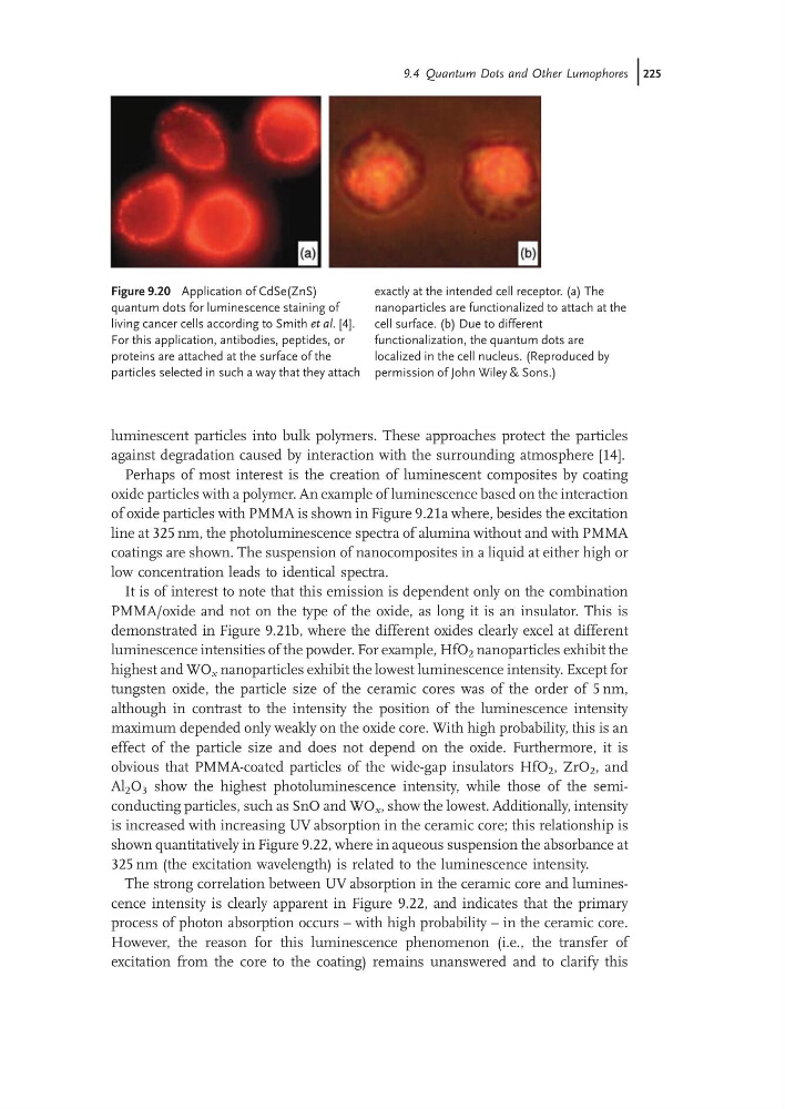
Figure 9.20 Application of CdSe(ZnS)
quantum dots for luminescence staining of
living cancer cells according to Smith et al. [4].
For this application, antibodies, peptides, or
proteins are attached at the surface of the
particles selected in such a way that they attach
exactly at the intended cell receptor. (a) The
nanoparticles are functionalized to attach at the
cell surface. (b) Due to different
functionalization, the quantum dots are
localized in the cell nucleus. (Reproduced by
permission of John Wiley & Sons.)
9.4 Quantum Dots and Other Lumophores j225
against degradation caused by interaction with the surrounding atmosphere [14].
Perhaps of most interest is the creation of luminescent composites by coating
oxide particles with a polymer. An example of luminescence based on the interaction
of oxide particles with PMMA is shown in Figure 9.21a where, besides the excitation
line at 325 nm, the photoluminescence spectra of alumina without and with PMMA
coatings are shown. The suspension of nanocomposites in a liquid at either high or
low concentration leads to identical spectra.
It is of interest to note that this emission is dependent only on the combination
PMMA/oxide and not on the type of the oxide, as long it is an insulator. This is
demonstrated in Figure 9.21b, where the different oxides clearly excel at different
luminescence intensities of the powder. For example, HfO 2 nanoparticles exhibit the
highest and WO x nanoparticles exhibit the lowest luminescence intensity. Except for
tungsten oxide, the particle size of the ceramic cores was of the order of 5 nm,
although in contrast to the intensity the position of the luminescence intensity
maximum depended only weakly on the oxide core. With high probability, this is an
effect of the particle size and does not depend on the oxide. Furthermore, it is
obvious that PMMA-coated particles of the wide-gap insulators HfO 2 , ZrO 2 , and
Al 2 O 3 show the highest photoluminescence intensity, while those of the semiconducting particles, such as SnO and WO x , show the lowest. Additionally, intensity
is increased with increasing UV absorption in the ceramic core; this relationship is
shown quantitatively in Figure 9.22, where in aqueous suspension the absorbance at
325 nm (the excitation wavelength) is related to the luminescence intensity.
The strong correlation between UV absorption in the ceramic core and luminescence intensity is clearly apparent in Figure 9.22, and indicates that the primary
process of photon absorption occurs – with high probability – in the ceramic core.
However, the reason for this luminescence phenomenon (i.e., the transfer of
excitation from the core to the coating) remains unanswered and to clarify this
Figure 9.20 Application of CdSe(ZnS)
quantum dots for luminescence staining of
living cancer cells according to Smith et al. [4].
For this application, antibodies, peptides, or
proteins are attached at the surface of the
particles selected in such a way that they attach
exactly at the intended cell receptor. (a) The
nanoparticles are functionalized to attach at the
cell surface. (b) Due to different
functionalization, the quantum dots are
localized in the cell nucleus. (Reproduced by
permission of John Wiley & Sons.)
9.4 Quantum Dots and Other Lumophores j225