

An example of nanoplates (in this case gold nanoplates) is shown in Figure 5.3a.
The size of these platelets is approximately 400 nm in the plane and their thickness
ranges from 25 to 60 nm. As can be seen from the hexagonal shape of the platelets,
the nanoplates have a single orientation with the [111] direction perpendicular to the
plane. This is also clearly visible in the electron diffraction pattern (Figure 5.3b),
which shows the [111] reflexes as the most intensive. This example is of special
interest because gold crystallizes in a cubic structure and therefore cubes rather than
platelets would be expected. However, the synthesis process applied in this case was
Figure 5.1 Secondary electron micrograph of
ZnO nanorods [1]. These nanorods have lengths
of about 15 mm and diameters ranging from 120
to 140 nm. At one end, most of the nanorods
show a, possibly metallic, bulge, which is typical
of synthesis via a gas-phase route. (Reproduced
by permission of Springer.)
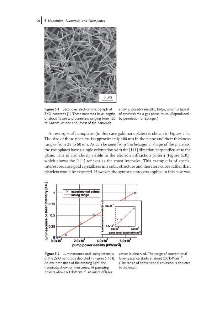
Figure 5.2 Luminescence and lasing intensity
of the ZnO nanorods depicted in Figure 5.1 [1].
At low intensities of the exciting light, the
nanorods show luminescence. At pumping
powers above 600 kW cm
À2
, an onset of laser
action is observed. The range of conventional
luminescence starts at about 200 kW cm
À2 .
(The range of conventional emission is depicted
in the inset.)
90j 5 Nanotubes, Nanorods, and Nanoplates
The size of these platelets is approximately 400 nm in the plane and their thickness
ranges from 25 to 60 nm. As can be seen from the hexagonal shape of the platelets,
the nanoplates have a single orientation with the [111] direction perpendicular to the
plane. This is also clearly visible in the electron diffraction pattern (Figure 5.3b),
which shows the [111] reflexes as the most intensive. This example is of special
interest because gold crystallizes in a cubic structure and therefore cubes rather than
platelets would be expected. However, the synthesis process applied in this case was
Figure 5.1 Secondary electron micrograph of
ZnO nanorods [1]. These nanorods have lengths
of about 15 mm and diameters ranging from 120
to 140 nm. At one end, most of the nanorods
show a, possibly metallic, bulge, which is typical
of synthesis via a gas-phase route. (Reproduced
by permission of Springer.)
Figure 5.2 Luminescence and lasing intensity
of the ZnO nanorods depicted in Figure 5.1 [1].
At low intensities of the exciting light, the
nanorods show luminescence. At pumping
powers above 600 kW cm
À2
, an onset of laser
action is observed. The range of conventional
luminescence starts at about 200 kW cm
À2 .
(The range of conventional emission is depicted
in the inset.)
90j 5 Nanotubes, Nanorods, and Nanoplates