

2.3 Characterisation Techniques
45
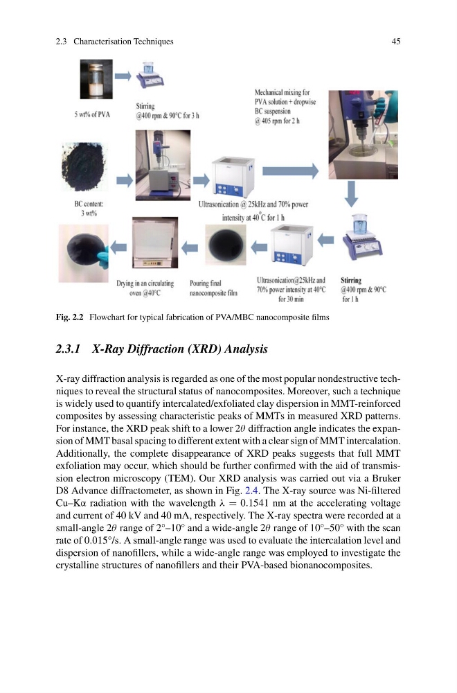
Fig. 2.2 Flowchart for typical fabrication of PVA/MBC nanocomposite films
2.3.1 X-Ray Diffraction (XRD) Analysis
X-ray diffraction analysis is regarded as one of the most popular nondestructive techniques to reveal the structural status of nanocomposites. Moreover, such a technique
is widely used to quantify intercalated/exfoliated clay dispersion in MMT-reinforced
composites by assessing characteristic peaks of MMTs in measured XRD patterns.
For instance, the XRD peak shift to a lower 2θ diffraction angle indicates the expansion of MMT basal spacing to different extent with a clear sign of MMT intercalation.
Additionally, the complete disappearance of XRD peaks suggests that full MMT
exfoliation may occur, which should be further confirmed with the aid of transmission electron microscopy (TEM). Our XRD analysis was carried out via a Bruker
D8 Advance diffractometer, as shown in Fig. 2.4. The X-ray source was Ni-filtered
Cu–Kα radiation with the wavelength λ = 0.1541 nm at the accelerating voltage
and current of 40 kV and 40 mA, respectively. The X-ray spectra were recorded at a
small-angle 2θ range of 2°–10° and a wide-angle 2θ range of 10°–50° with the scan
rate of 0.015°/s. A small-angle range was used to evaluate the intercalation level and
dispersion of nanofillers, while a wide-angle range was employed to investigate the
crystalline structures of nanofillers and their PVA-based bionanocomposites.
45
Fig. 2.2 Flowchart for typical fabrication of PVA/MBC nanocomposite films
2.3.1 X-Ray Diffraction (XRD) Analysis
X-ray diffraction analysis is regarded as one of the most popular nondestructive techniques to reveal the structural status of nanocomposites. Moreover, such a technique
is widely used to quantify intercalated/exfoliated clay dispersion in MMT-reinforced
composites by assessing characteristic peaks of MMTs in measured XRD patterns.
For instance, the XRD peak shift to a lower 2θ diffraction angle indicates the expansion of MMT basal spacing to different extent with a clear sign of MMT intercalation.
Additionally, the complete disappearance of XRD peaks suggests that full MMT
exfoliation may occur, which should be further confirmed with the aid of transmission electron microscopy (TEM). Our XRD analysis was carried out via a Bruker
D8 Advance diffractometer, as shown in Fig. 2.4. The X-ray source was Ni-filtered
Cu–Kα radiation with the wavelength λ = 0.1541 nm at the accelerating voltage
and current of 40 kV and 40 mA, respectively. The X-ray spectra were recorded at a
small-angle 2θ range of 2°–10° and a wide-angle 2θ range of 10°–50° with the scan
rate of 0.015°/s. A small-angle range was used to evaluate the intercalation level and
dispersion of nanofillers, while a wide-angle range was employed to investigate the
crystalline structures of nanofillers and their PVA-based bionanocomposites.