

4.6 Aspect Ratios of Embedded Nanofillers in PVA Bionanocomposites
93
A16
B16
A15
B15
A16
A15
B15
B16
(b)
(a)
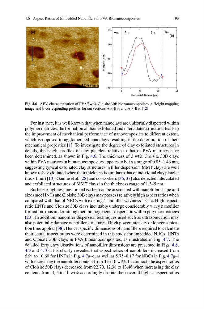
Fig. 4.6 AFM characterisation of PVA/3wt% Cloisite 30B bionanocomposites. a Height mapping
image and b corresponding profiles for cut sections A 15 -B 15 and A 16 -B 16 [12]
For instance, it is well known that when nanoclays are uniformly dispersed within
polymer matrices, the formation of their exfoliated and intercalated structures leads to
the improvement of mechanical performance of nanocomposites to different extent,
which is opposed to agglomerated nanoclays resulting in the deterioration of their
mechanical properties [1]. To investigate the degree of clay exfoliated structures in
details, the height profiles of clay platelets relative to that of PVA matrices have
been determined, as shown in Fig. 4.6. The thickness of 3 wt% Cloisite 30B clays
within PVA matrices in bionanocomposites appears to be in a range of 0.85–1.43 nm,
suggesting typical exfoliated clay structures in filler dispersion. MMT clays are well
known to be exfoliated when their thickness is similar to that of individual clay platelet
(i.e. ~1 nm) [13]. Gaume et al. [28] and co-workers [36, 37] also detected intercalated
and exfoliated structures of MMT clays in the thickness range of 1.3–5 nm.
Surface roughness mentioned earlier can be associated with nanofiller shape and
size since HNTs and Cloisite 30B clays may possess relatively high aspect ratios when
compared with that of NBCs with existing ‘nanofiller waviness’ issue. High-aspectratio HNTs and Cloisite 30B clays inevitably undergo considerably wavy nanofiller
formation, thus undermining their homogeneous dispersion within polymer matrices
[23]. In addition, nanofiller dispersion techniques used such as ultrasonication may
also potentially damage nanofiller structures if high power intensity or longer sonication time applies [38]. Hence, specific dimensions of nanofillers required to calculate
their actual aspect ratios were determined in this study for embedded NBCs, HNTs
and Cloisite 30B clays in PVA bionanocomposites, as illustrated in Fig. 4.7. The
detailed frequency distributions of nanofiller dimensions are presented in Figs. 4.8,
4.9 and 4.10. It is clearly revealed that aspect ratios of nanofillers increased from
5.91 to 10.60 for HNTs in Fig. 4.7a–c, as well as 5.75–8.17 for NBCs in Fig. 4.7g–i
with increasing the nanofiller content from 3 to 10 wt%. In contrast, the aspect ratios
of Cloisite 30B clays decreased from 22.70, 12.38 to 13.46 when increasing the clay
contents from 3, 5 to 10 wt% accordingly despite their overall highest aspect ratios
93
A16
B16
A15
B15
A16
A15
B15
B16
(b)
(a)
Fig. 4.6 AFM characterisation of PVA/3wt% Cloisite 30B bionanocomposites. a Height mapping
image and b corresponding profiles for cut sections A 15 -B 15 and A 16 -B 16 [12]
For instance, it is well known that when nanoclays are uniformly dispersed within
polymer matrices, the formation of their exfoliated and intercalated structures leads to
the improvement of mechanical performance of nanocomposites to different extent,
which is opposed to agglomerated nanoclays resulting in the deterioration of their
mechanical properties [1]. To investigate the degree of clay exfoliated structures in
details, the height profiles of clay platelets relative to that of PVA matrices have
been determined, as shown in Fig. 4.6. The thickness of 3 wt% Cloisite 30B clays
within PVA matrices in bionanocomposites appears to be in a range of 0.85–1.43 nm,
suggesting typical exfoliated clay structures in filler dispersion. MMT clays are well
known to be exfoliated when their thickness is similar to that of individual clay platelet
(i.e. ~1 nm) [13]. Gaume et al. [28] and co-workers [36, 37] also detected intercalated
and exfoliated structures of MMT clays in the thickness range of 1.3–5 nm.
Surface roughness mentioned earlier can be associated with nanofiller shape and
size since HNTs and Cloisite 30B clays may possess relatively high aspect ratios when
compared with that of NBCs with existing ‘nanofiller waviness’ issue. High-aspectratio HNTs and Cloisite 30B clays inevitably undergo considerably wavy nanofiller
formation, thus undermining their homogeneous dispersion within polymer matrices
[23]. In addition, nanofiller dispersion techniques used such as ultrasonication may
also potentially damage nanofiller structures if high power intensity or longer sonication time applies [38]. Hence, specific dimensions of nanofillers required to calculate
their actual aspect ratios were determined in this study for embedded NBCs, HNTs
and Cloisite 30B clays in PVA bionanocomposites, as illustrated in Fig. 4.7. The
detailed frequency distributions of nanofiller dimensions are presented in Figs. 4.8,
4.9 and 4.10. It is clearly revealed that aspect ratios of nanofillers increased from
5.91 to 10.60 for HNTs in Fig. 4.7a–c, as well as 5.75–8.17 for NBCs in Fig. 4.7g–i
with increasing the nanofiller content from 3 to 10 wt%. In contrast, the aspect ratios
of Cloisite 30B clays decreased from 22.70, 12.38 to 13.46 when increasing the clay
contents from 3, 5 to 10 wt% accordingly despite their overall highest aspect ratios