

standard gentamicin protection assay [10]. Visualizing internalized
bacteria and its progress within cells using confocal microscopy
supplies a much clearer picture of vectors’ abilities to successfully
internalize into the cytoplasm (Fig. 2), but is not used routinely
when studying molecule delivery using bacteria.
Here, we demonstrate the use of high-resolution microscopy
including Scanning Electron Microscopy (SEM) and confocal
microscopy to visualize the attachment and internalization characteristics of invasive E. coli during the invasion of human cells. These
techniques can be easily and cost-effectively adapted for analysis of
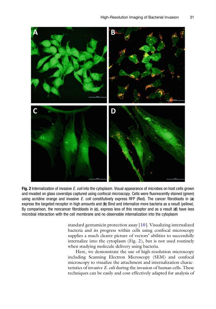
Fig. 2 Internalization of invasive E. coli into the cytoplasm. Visual appearance of microbes on host cells grown
and invaded on glass coverslips captured using confocal microscopy. Cells were fluorescently stained (green)
using acridine orange and invasive E. coli constitutively express RFP (Red). The cancer fibroblasts in (a)
express the targeted receptor in high amounts and (b) Bind and internalize more bacteria as a result (yellow).
By comparison, the noncancer fibroblasts in (c), express less of this receptor and as a result (d) have less
microbial interaction with the cell membrane and no observable internalization into the cytoplasm
High-Resolution Imaging of Bacterial Invasion
31
bacteria and its progress within cells using confocal microscopy
supplies a much clearer picture of vectors’ abilities to successfully
internalize into the cytoplasm (Fig. 2), but is not used routinely
when studying molecule delivery using bacteria.
Here, we demonstrate the use of high-resolution microscopy
including Scanning Electron Microscopy (SEM) and confocal
microscopy to visualize the attachment and internalization characteristics of invasive E. coli during the invasion of human cells. These
techniques can be easily and cost-effectively adapted for analysis of
Fig. 2 Internalization of invasive E. coli into the cytoplasm. Visual appearance of microbes on host cells grown
and invaded on glass coverslips captured using confocal microscopy. Cells were fluorescently stained (green)
using acridine orange and invasive E. coli constitutively express RFP (Red). The cancer fibroblasts in (a)
express the targeted receptor in high amounts and (b) Bind and internalize more bacteria as a result (yellow).
By comparison, the noncancer fibroblasts in (c), express less of this receptor and as a result (d) have less
microbial interaction with the cell membrane and no observable internalization into the cytoplasm
High-Resolution Imaging of Bacterial Invasion
31