

9. Transform pCYC-LyE by electroporation into select E. coli
bactofection strains. In this case, the plasmid was electroporated into strains BL21(DE3)/pET29-hly/pCMV-Luc,
YWT7-hly/pCMV-Luc, and YWTet-hly/pCMV-Luc using a
MicroPulser electroporator (see Note 3).
3.2 Evaluating
Bacterial Growth
Inhibition by LyE
1. Inoculate E. coli strains into 3 mL of lysogeny broth (LB) medium containing the appropriate antibiotics within an orbital
shaker at 250 rpm and 37
C and grow overnight for 16–20 h
(see Note 4).
2. Transfer 1.5 mL of the seed culture into a 200 mL flask containing 50 mL of fresh LB media with the appropriate antibiotics and culture under the same conditions.
3. Quantify the cell density within the culture by measuring the
optical density at 600 nm (OD 600nm ) using a GENEYS 20 visible spectrophotometer. Continue taking measurements until
an OD 600nm of 0.4–0.5 is reached (see Note 5).
4. Once an OD 600nm of 0.4–0.5 has been reached, add isopropyl
β-D-1-thiogalactopyranoside (IPTG) at varying concentrations
(0, 100, 500, 1000, 2000 μM) to introduce gene expression.
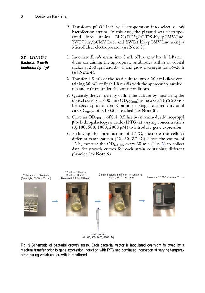
5. Following the introduction of IPTG, incubate the cells at
different temperatures (22, 30, 37
C). Over the course of
12 h, measure the OD 600nm every 30 min (Fig. 3) to collect
data for growth curves for each strain containing different
plasmids (see Note 6).
Culture 3 mL of bacteria
(Overnight, 36 °C, 250 rpm)
1.5 mL of culture in
50 mL of LB broth
(Overnight, 36 °C, 250 rpm)
Culture bacteria in different temperature:
(22, 30, 37 °C, 250 rpm)
Measure OD 600nm every 30 min
IPTG injection
(0, 100, 500, 1000, 2000 µM)
Fig. 3 Schematic of bacterial growth assay. Each bacterial vector is inoculated overnight followed by a
medium transfer prior to gene expression induction with IPTG and continued incubation at varying temperatures during which cell growth is monitored
8
Dongwon Park et al.
bactofection strains. In this case, the plasmid was electroporated into strains BL21(DE3)/pET29-hly/pCMV-Luc,
YWT7-hly/pCMV-Luc, and YWTet-hly/pCMV-Luc using a
MicroPulser electroporator (see Note 3).
3.2 Evaluating
Bacterial Growth
Inhibition by LyE
1. Inoculate E. coli strains into 3 mL of lysogeny broth (LB) medium containing the appropriate antibiotics within an orbital
shaker at 250 rpm and 37
C and grow overnight for 16–20 h
(see Note 4).
2. Transfer 1.5 mL of the seed culture into a 200 mL flask containing 50 mL of fresh LB media with the appropriate antibiotics and culture under the same conditions.
3. Quantify the cell density within the culture by measuring the
optical density at 600 nm (OD 600nm ) using a GENEYS 20 visible spectrophotometer. Continue taking measurements until
an OD 600nm of 0.4–0.5 is reached (see Note 5).
4. Once an OD 600nm of 0.4–0.5 has been reached, add isopropyl
β-D-1-thiogalactopyranoside (IPTG) at varying concentrations
(0, 100, 500, 1000, 2000 μM) to introduce gene expression.
5. Following the introduction of IPTG, incubate the cells at
different temperatures (22, 30, 37
C). Over the course of
12 h, measure the OD 600nm every 30 min (Fig. 3) to collect
data for growth curves for each strain containing different
plasmids (see Note 6).
Culture 3 mL of bacteria
(Overnight, 36 °C, 250 rpm)
1.5 mL of culture in
50 mL of LB broth
(Overnight, 36 °C, 250 rpm)
Culture bacteria in different temperature:
(22, 30, 37 °C, 250 rpm)
Measure OD 600nm every 30 min
IPTG injection
(0, 100, 500, 1000, 2000 µM)
Fig. 3 Schematic of bacterial growth assay. Each bacterial vector is inoculated overnight followed by a
medium transfer prior to gene expression induction with IPTG and continued incubation at varying temperatures during which cell growth is monitored
8
Dongwon Park et al.