

synthetic counterparts owing to their autologous sources, little is
known about their long-term immune stimulation and immunogenicity potential. Furthermore, the shearing process of CDNs production, while retaining crucial protein markers of endogenously
produced exosomes, may potentially alter the membrane conformation and composition [38]. Nonetheless, further studies are
necessary to identify the specific components responsible for the
high organotropism of exosomes. Moreover, future development
of exosome-mimetics aimed to overcome the drawbacks of the
current DDSs will pave the way for the development of an ultimate
safe and targeted delivery system.
In the following sections, we cover the step-by-step procedures
for producing CDNs as reported by Goh et al. [29]. The rationale
for each step is discussed in the “Notes” sections.
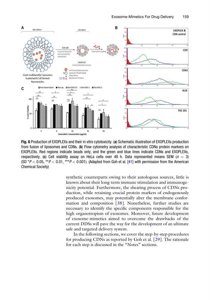
Fig. 6 Production of EXOPLEXs and their in vitro cytotoxicity. (a) Schematic illustration of EXOPLEXs production
from fusion of liposomes and CDNs. (b) Flow cytometry analysis of characteristic CDNs protein markers on
EXOPLEXs. Red regions indicate beads only, and the green and blue lines indicate CDNs and EXOPLEXs,
respectively. (c) Cell viability assay on HeLa cells over 48 h. Data represented means SEM (n ¼ 3)
(SD *P < 0.05, **P < 0.01, ***P < 0.001). (Adapted from Goh et al. [41] with permission from the American
Chemical Society)
Exosome-Mimetics For Drug Delivery
159
known about their long-term immune stimulation and immunogenicity potential. Furthermore, the shearing process of CDNs production, while retaining crucial protein markers of endogenously
produced exosomes, may potentially alter the membrane conformation and composition [38]. Nonetheless, further studies are
necessary to identify the specific components responsible for the
high organotropism of exosomes. Moreover, future development
of exosome-mimetics aimed to overcome the drawbacks of the
current DDSs will pave the way for the development of an ultimate
safe and targeted delivery system.
In the following sections, we cover the step-by-step procedures
for producing CDNs as reported by Goh et al. [29]. The rationale
for each step is discussed in the “Notes” sections.
Fig. 6 Production of EXOPLEXs and their in vitro cytotoxicity. (a) Schematic illustration of EXOPLEXs production
from fusion of liposomes and CDNs. (b) Flow cytometry analysis of characteristic CDNs protein markers on
EXOPLEXs. Red regions indicate beads only, and the green and blue lines indicate CDNs and EXOPLEXs,
respectively. (c) Cell viability assay on HeLa cells over 48 h. Data represented means SEM (n ¼ 3)
(SD *P < 0.05, **P < 0.01, ***P < 0.001). (Adapted from Goh et al. [41] with permission from the American
Chemical Society)
Exosome-Mimetics For Drug Delivery
159