

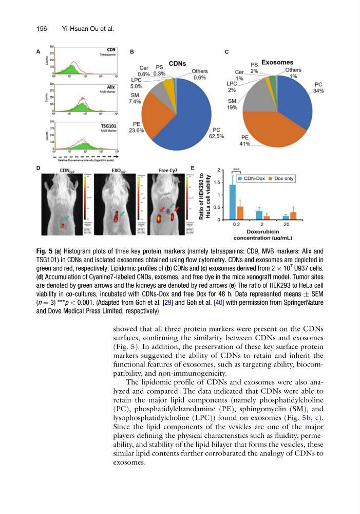
showed that all three protein markers were present on the CDNs
surfaces, confirming the similarity between CDNs and exosomes
(Fig. 5). In addition, the preservation of these key surface protein
markers suggested the ability of CDNs to retain and inherit the
functional features of exosomes, such as targeting ability, biocompatibility, and non-immunogenicity.
The lipidomic profile of CDNs and exosomes were also analyzed and compared. The data indicated that CDNs were able to
retain the major lipid components (namely phosphatidylcholine
(PC), phosphatidylehanolamine (PE), sphingomyelin (SM), and
lysophosphatidylcholine (LPC)) found on exosomes (Fig. 5b, c).
Since the lipid components of the vesicles are one of the major
players defining the physical characteristics such as fluidity, permeability, and stability of the lipid bilayer that forms the vesicles, these
similar lipid contents further corrobarated the analogy of CDNs to
exosomes.
Fig. 5 (a) Histogram plots of three key protein markers (namely tetraspanins: CD9, MVB markers: Alix and
TSG101) in CDNs and isolated exosomes obtained using flow cytometry. CDNs and exosomes are depicted in
green and red, respectively. Lipidomic profiles of (b) CDNs and (c) exosomes derived from 2 Â 10
7
U937 cells.
(d) Accumulation of Cyanine7-labeled CNDs, exosmes, and free dye in the mice xenograft model. Tumor sites
are denoted by green arrows and the kidneys are denoted by red arrows (e) The ratio of HEK293 to HeLa cell
viability in co-cultures, incubated with CDNs-Dox and free Dox for 48 h. Data represented means Æ SEM
(n ¼ 3) ***p < 0.001. (Adapted from Goh et al. [29] and Goh et al. [40] with permission from SpringerNature
and Dove Medical Press Limited, respectively)
156
Yi-Hsuan Ou et al.
surfaces, confirming the similarity between CDNs and exosomes
(Fig. 5). In addition, the preservation of these key surface protein
markers suggested the ability of CDNs to retain and inherit the
functional features of exosomes, such as targeting ability, biocompatibility, and non-immunogenicity.
The lipidomic profile of CDNs and exosomes were also analyzed and compared. The data indicated that CDNs were able to
retain the major lipid components (namely phosphatidylcholine
(PC), phosphatidylehanolamine (PE), sphingomyelin (SM), and
lysophosphatidylcholine (LPC)) found on exosomes (Fig. 5b, c).
Since the lipid components of the vesicles are one of the major
players defining the physical characteristics such as fluidity, permeability, and stability of the lipid bilayer that forms the vesicles, these
similar lipid contents further corrobarated the analogy of CDNs to
exosomes.
Fig. 5 (a) Histogram plots of three key protein markers (namely tetraspanins: CD9, MVB markers: Alix and
TSG101) in CDNs and isolated exosomes obtained using flow cytometry. CDNs and exosomes are depicted in
green and red, respectively. Lipidomic profiles of (b) CDNs and (c) exosomes derived from 2 Â 10
7
U937 cells.
(d) Accumulation of Cyanine7-labeled CNDs, exosmes, and free dye in the mice xenograft model. Tumor sites
are denoted by green arrows and the kidneys are denoted by red arrows (e) The ratio of HEK293 to HeLa cell
viability in co-cultures, incubated with CDNs-Dox and free Dox for 48 h. Data represented means Æ SEM
(n ¼ 3) ***p < 0.001. (Adapted from Goh et al. [29] and Goh et al. [40] with permission from SpringerNature
and Dove Medical Press Limited, respectively)
156
Yi-Hsuan Ou et al.