

analyses to demonstrated that PNAs are internalized within
target cells. Confocal analyses might in addition be very useful
if PNA distribution within intracellular compartments should
be determined, as pointed out in Brognara et al. [46].
18. It should be emphasized that the binding of PNA to microRNA does not alter the production of miRNA, and that there is
no evidence so far that PNA can induce degradation of the
target microRNA. Thus, the measurements performed using
RT-qPCR do not measure the microRNA expression, but
rather indicate its bioavailability, which is dependent on the
amount of free microRNA which is not sequestered by PNA.
For a discussion of the cellular fate of microRNA after
PNA/LNA binding see Torres et al. [74].
19. As for specificity on PNA-mediated miRNA inhibition (verified
quantifying free miRNAs using RT-qPCR), the results
obtained demonstrate that first of all culturing U251 cells in
the presence of the PNA-a221 (lacking the R8 peptide) is not
associated with miR-221-3p inhibition (Fig. 3a). On the contrary, miR-221-3p hybridization signals are deeply inhibited
when the U251 cells have been cultured in the presence of
R8-PNA-a221. Interestingly, the incubation of U251 cells
with the argininocalix[4]arene 1/PNA-a221 formulation
leads to results very similar to that obtained with the
R8-PNA-a221 and fully compatible with the concept that
high level of cellular uptake is obtained by the delivery of the
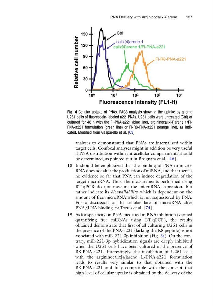
Fig. 4 Cellular uptake of PNAs. FACS analysis showing the uptake by glioma
U251 cells of fluorescein-labeled a221PNAs. U251 cells were untreated (Ctrl) or
cultured for 48 h with the Fl-PNA-a221 (blue line), argininocalix[4]arene 1/FlPNA-a221 formulation (green line) or Fl-R8-PNA-a221 (orange line), as indicated. Modified from Gasparello et al. [63]
PNA Delivery with Argininocalix[4]arene
137
target cells. Confocal analyses might in addition be very useful
if PNA distribution within intracellular compartments should
be determined, as pointed out in Brognara et al. [46].
18. It should be emphasized that the binding of PNA to microRNA does not alter the production of miRNA, and that there is
no evidence so far that PNA can induce degradation of the
target microRNA. Thus, the measurements performed using
RT-qPCR do not measure the microRNA expression, but
rather indicate its bioavailability, which is dependent on the
amount of free microRNA which is not sequestered by PNA.
For a discussion of the cellular fate of microRNA after
PNA/LNA binding see Torres et al. [74].
19. As for specificity on PNA-mediated miRNA inhibition (verified
quantifying free miRNAs using RT-qPCR), the results
obtained demonstrate that first of all culturing U251 cells in
the presence of the PNA-a221 (lacking the R8 peptide) is not
associated with miR-221-3p inhibition (Fig. 3a). On the contrary, miR-221-3p hybridization signals are deeply inhibited
when the U251 cells have been cultured in the presence of
R8-PNA-a221. Interestingly, the incubation of U251 cells
with the argininocalix[4]arene 1/PNA-a221 formulation
leads to results very similar to that obtained with the
R8-PNA-a221 and fully compatible with the concept that
high level of cellular uptake is obtained by the delivery of the
Fig. 4 Cellular uptake of PNAs. FACS analysis showing the uptake by glioma
U251 cells of fluorescein-labeled a221PNAs. U251 cells were untreated (Ctrl) or
cultured for 48 h with the Fl-PNA-a221 (blue line), argininocalix[4]arene 1/FlPNA-a221 formulation (green line) or Fl-R8-PNA-a221 (orange line), as indicated. Modified from Gasparello et al. [63]
PNA Delivery with Argininocalix[4]arene
137