

3.2.2 Fluorescent
Microscopy
1. Detect the EGFP reporter gene expression using an epifluorescent or confocal microscope equipped with a digital or
cooled charge-coupled device (CCD) camera.
2. Record bright-field images for cell morphology.
3. Capture green fluorescent images under GFP channel for the
EGFP reporter gene expression in cells (see Note 10).
4 Notes
1. Agarose gel solution should be handled with caution during
microwave heating as explosive boiling is a hazard. Agarose gel
solution should be heated in a microwave oven for a few
seconds/minutes and heating should be stopped right before
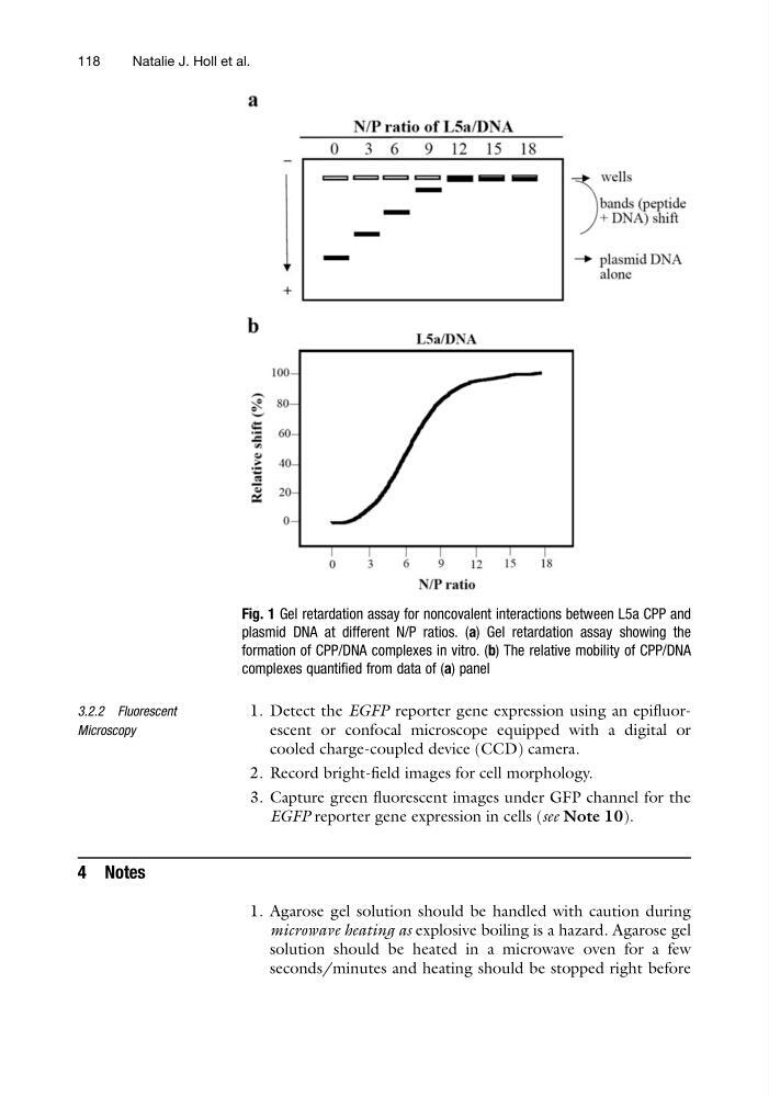
Fig. 1 Gel retardation assay for noncovalent interactions between L5a CPP and
plasmid DNA at different N/P ratios. (a) Gel retardation assay showing the
formation of CPP/DNA complexes in vitro. (b) The relative mobility of CPP/DNA
complexes quantified from data of (a) panel
118
Natalie J. Holl et al.
Microscopy
1. Detect the EGFP reporter gene expression using an epifluorescent or confocal microscope equipped with a digital or
cooled charge-coupled device (CCD) camera.
2. Record bright-field images for cell morphology.
3. Capture green fluorescent images under GFP channel for the
EGFP reporter gene expression in cells (see Note 10).
4 Notes
1. Agarose gel solution should be handled with caution during
microwave heating as explosive boiling is a hazard. Agarose gel
solution should be heated in a microwave oven for a few
seconds/minutes and heating should be stopped right before
Fig. 1 Gel retardation assay for noncovalent interactions between L5a CPP and
plasmid DNA at different N/P ratios. (a) Gel retardation assay showing the
formation of CPP/DNA complexes in vitro. (b) The relative mobility of CPP/DNA
complexes quantified from data of (a) panel
118
Natalie J. Holl et al.