

9 Mössbauer Spectroscopic Studies on Atomic Diffusion in Materials
475
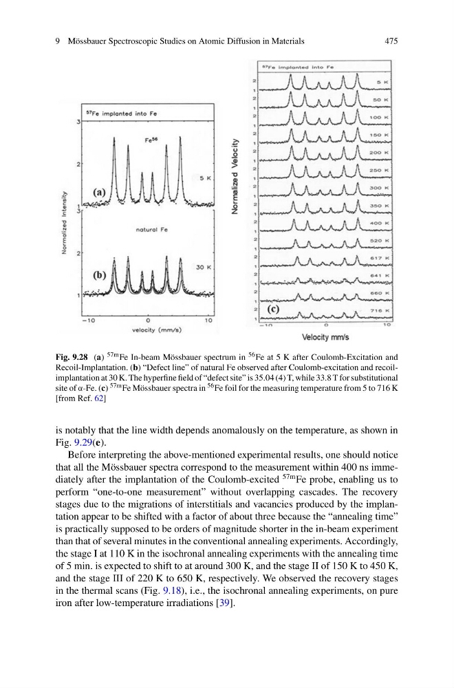
Fig. 9.28 (a) 57m Fe In-beam Mössbauer spectrum in 56 Fe at 5 K after Coulomb-Excitation and
Recoil-Implantation. (b) “Defect line” of natural Fe observed after Coulomb-excitation and recoilimplantation at 30 K. The hyperfine field of “defect site” is 35.04 (4) T, while 33.8 T for substitutional
site of α-Fe. (c) 57m Fe Mössbauer spectra in 56 Fe foil for the measuring temperature from 5 to 716 K
[from Ref. 62]
is notably that the line width depends anomalously on the temperature, as shown in
Fig. 9.29(e).
Before interpreting the above-mentioned experimental results, one should notice
that all the Mössbauer spectra correspond to the measurement within 400 ns immediately after the implantation of the Coulomb-excited
57m Fe probe, enabling us to
perform “one-to-one measurement” without overlapping cascades. The recovery
stages due to the migrations of interstitials and vacancies produced by the implantation appear to be shifted with a factor of about three because the “annealing time”
is practically supposed to be orders of magnitude shorter in the in-beam experiment
than that of several minutes in the conventional annealing experiments. Accordingly,
the stage I at 110 K in the isochronal annealing experiments with the annealing time
of 5 min. is expected to shift to at around 300 K, and the stage II of 150 K to 450 K,
and the stage III of 220 K to 650 K, respectively. We observed the recovery stages
in the thermal scans (Fig. 9.18), i.e., the isochronal annealing experiments, on pure
iron after low-temperature irradiations [39].
475
Fig. 9.28 (a) 57m Fe In-beam Mössbauer spectrum in 56 Fe at 5 K after Coulomb-Excitation and
Recoil-Implantation. (b) “Defect line” of natural Fe observed after Coulomb-excitation and recoilimplantation at 30 K. The hyperfine field of “defect site” is 35.04 (4) T, while 33.8 T for substitutional
site of α-Fe. (c) 57m Fe Mössbauer spectra in 56 Fe foil for the measuring temperature from 5 to 716 K
[from Ref. 62]
is notably that the line width depends anomalously on the temperature, as shown in
Fig. 9.29(e).
Before interpreting the above-mentioned experimental results, one should notice
that all the Mössbauer spectra correspond to the measurement within 400 ns immediately after the implantation of the Coulomb-excited
57m Fe probe, enabling us to
perform “one-to-one measurement” without overlapping cascades. The recovery
stages due to the migrations of interstitials and vacancies produced by the implantation appear to be shifted with a factor of about three because the “annealing time”
is practically supposed to be orders of magnitude shorter in the in-beam experiment
than that of several minutes in the conventional annealing experiments. Accordingly,
the stage I at 110 K in the isochronal annealing experiments with the annealing time
of 5 min. is expected to shift to at around 300 K, and the stage II of 150 K to 450 K,
and the stage III of 220 K to 650 K, respectively. We observed the recovery stages
in the thermal scans (Fig. 9.18), i.e., the isochronal annealing experiments, on pure
iron after low-temperature irradiations [39].