

4 From Small Molecules to Complex Systems: A Survey of Chemical …
205
4.5.1 Identification of Fe–S-Centers by Mössbauer
Spectroscopy
In collaborations with the laboratory of Alfred X. Trautwein and colleagues from
biology and biochemistry we have contributed to this field by the characterization
of rubredoxin-type proteins both with the iron in the Fe
2+ –S 4 and Fe
3+ –S 4 state
[81], the identification of a 2Fe–2S center in the FhuF protein which is part of
a plant siderophore-reductase system [82], and the identification of unusual 4Fe–
4S proteins with non-sulfur ligands like the radical S-adenosylmethionine enzyme
coporphyrinogen III oxidase HemN [83]. High-field Mössbauer spectroscopy also
helped to identify two essential diamagnetic 4Fe–4S centers in the evolutionarily
highly conserved ATP-binding cassette protein ABCE1 [84].
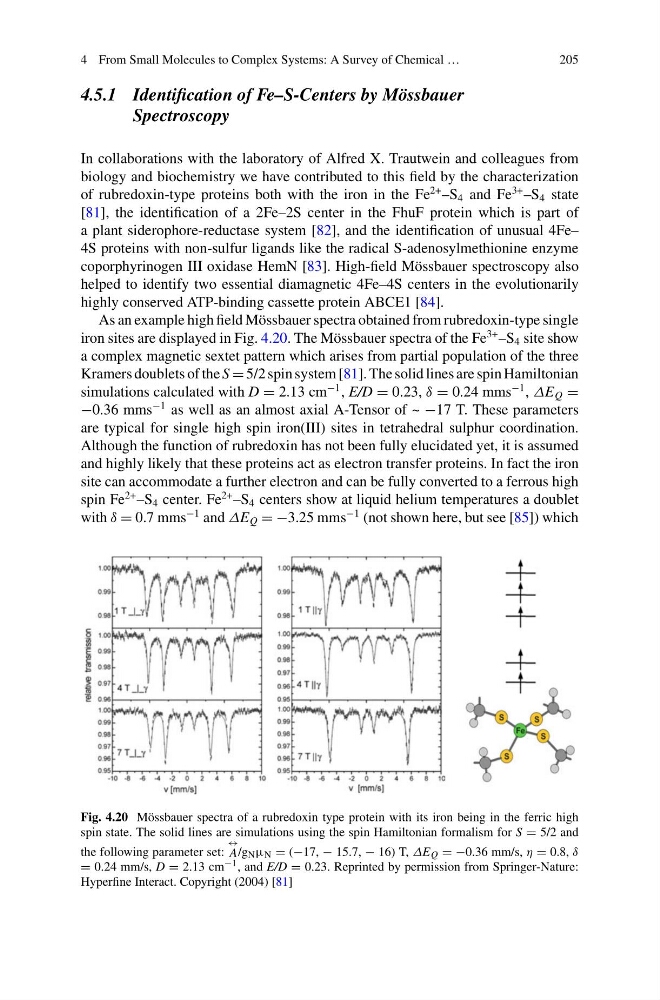
As an example high field Mössbauer spectra obtained from rubredoxin-type single
iron sites are displayed in Fig. 4.20. The Mössbauer spectra of the Fe
3+ –S 4 site show
a complex magnetic sextet pattern which arises from partial population of the three
Kramers doublets of the S = 5/2 spin system [81]. The solid lines are spin Hamiltonian
simulations calculated with D = 2.13 cm
−1 , E/D = 0.23, δ = 0.24 mms
−1 , ΔE Q =
−0.36 mms
−1 as well as an almost axial A-Tensor of ~ −17 T. These parameters
are typical for single high spin iron(III) sites in tetrahedral sulphur coordination.
Although the function of rubredoxin has not been fully elucidated yet, it is assumed
and highly likely that these proteins act as electron transfer proteins. In fact the iron
site can accommodate a further electron and can be fully converted to a ferrous high
spin Fe
2+ –S 4 center. Fe
2+ –S 4 centers show at liquid helium temperatures a doublet
with δ = 0.7 mms
−1 and ΔE Q = −3.25 mms
−1 (not shown here, but see [85]) which
Fig. 4.20 Mössbauer spectra of a rubredoxin type protein with its iron being in the ferric high
spin state. The solid lines are simulations using the spin Hamiltonian formalism for S = 5/2 and
the following parameter set:
↔
A/gNμN = (−17, − 15.7, − 16) T, ΔE Q = −0.36 mm/s, η = 0.8, δ
= 0.24 mm/s, D = 2.13 cm −1 , and E/D = 0.23. Reprinted by permission from Springer-Nature:
Hyperfine Interact. Copyright (2004) [81]
205
4.5.1 Identification of Fe–S-Centers by Mössbauer
Spectroscopy
In collaborations with the laboratory of Alfred X. Trautwein and colleagues from
biology and biochemistry we have contributed to this field by the characterization
of rubredoxin-type proteins both with the iron in the Fe
2+ –S 4 and Fe
3+ –S 4 state
[81], the identification of a 2Fe–2S center in the FhuF protein which is part of
a plant siderophore-reductase system [82], and the identification of unusual 4Fe–
4S proteins with non-sulfur ligands like the radical S-adenosylmethionine enzyme
coporphyrinogen III oxidase HemN [83]. High-field Mössbauer spectroscopy also
helped to identify two essential diamagnetic 4Fe–4S centers in the evolutionarily
highly conserved ATP-binding cassette protein ABCE1 [84].
As an example high field Mössbauer spectra obtained from rubredoxin-type single
iron sites are displayed in Fig. 4.20. The Mössbauer spectra of the Fe
3+ –S 4 site show
a complex magnetic sextet pattern which arises from partial population of the three
Kramers doublets of the S = 5/2 spin system [81]. The solid lines are spin Hamiltonian
simulations calculated with D = 2.13 cm
−1 , E/D = 0.23, δ = 0.24 mms
−1 , ΔE Q =
−0.36 mms
−1 as well as an almost axial A-Tensor of ~ −17 T. These parameters
are typical for single high spin iron(III) sites in tetrahedral sulphur coordination.
Although the function of rubredoxin has not been fully elucidated yet, it is assumed
and highly likely that these proteins act as electron transfer proteins. In fact the iron
site can accommodate a further electron and can be fully converted to a ferrous high
spin Fe
2+ –S 4 center. Fe
2+ –S 4 centers show at liquid helium temperatures a doublet
with δ = 0.7 mms
−1 and ΔE Q = −3.25 mms
−1 (not shown here, but see [85]) which
Fig. 4.20 Mössbauer spectra of a rubredoxin type protein with its iron being in the ferric high
spin state. The solid lines are simulations using the spin Hamiltonian formalism for S = 5/2 and
the following parameter set:
↔
A/gNμN = (−17, − 15.7, − 16) T, ΔE Q = −0.36 mm/s, η = 0.8, δ
= 0.24 mm/s, D = 2.13 cm −1 , and E/D = 0.23. Reprinted by permission from Springer-Nature:
Hyperfine Interact. Copyright (2004) [81]