

198
V. Schünemann
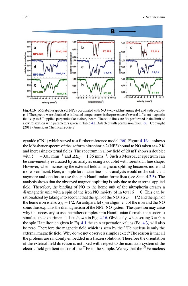
Fig. 4.16 Mössbauer spectra of NP2 coordinated with NO a–c, with histamine d–f and with cyanide
g–i. The spectra were obtained at indicated temperatures in the presence of several different magnetic
fields up to 5 T applied perpendicular to the γ-beam. The solid lines are fits performed in the limit of
slow relaxation with parameters given in Table 4.1. Adapted with permission from [66]. Copyright
(2012) American Chemical Society
cyanide (CN
− ) which served as a further reference model [66]. Figure 4.16a–c shows
the Mössbauer spectra of the isoform nitrophorin 2 (NP2) bound to NO taken at 4.2 K
and increasing external fields. The spectrum in a low field of 20 mT shows a doublet
with δ = −0.01 mms
−1 and ΔE Q = 1.86 mms
−1 . Such a Mössbauer spectrum can
be conveniently evaluated by an analysis using a doublet with lorentzian line shape.
However, when increasing the external field a magnetic splitting becomes more and
more prominent. Here, a simple lorentzian line shape analysis would not be sufficient
anymore and one has to use the spin Hamiltonian formalism (see Sect. 4.2.3). The
analysis shows that the observed magnetic splitting is only due to the external applied
field. Therefore, the binding of NO to the heme unit of the nitrophorin creates a
diamagnetic unit with a spin of the iron NO moiety of in total S = 0. This can be
rationalized by taking into account that the spin of the NO is S NO = 1/2 and the spin of
the heme iron is also S Fe = 1/2. An antiparallel spin alignment of the iron and the NO
spins thus explains the diamagnetism of the NP2–NO system. The question may arise
why it is necessary to use the rather complex spin Hamiltonian formalism in order to
simulate the experimental data shown in Fig. 4.16. Obviously, when setting S = 0 in
the spin Hamiltonian given in Eq. 4.1 the spin expectation values (Eq. 4.3) will also
be zero. Therefore the magnetic field which is seen by the
57 Fe nucleus is only the
external magnetic field. Why do we not observe a simple sextet? The reason is that all
the proteins are randomly embedded in a frozen solutions. Therefore the orientation
of the external field direction is not fixed with respect to the main axis system of the
electric field gradient tensor of the
57 Fe in the sample. We say that the
57 Fe nucleus
V. Schünemann
Fig. 4.16 Mössbauer spectra of NP2 coordinated with NO a–c, with histamine d–f and with cyanide
g–i. The spectra were obtained at indicated temperatures in the presence of several different magnetic
fields up to 5 T applied perpendicular to the γ-beam. The solid lines are fits performed in the limit of
slow relaxation with parameters given in Table 4.1. Adapted with permission from [66]. Copyright
(2012) American Chemical Society
cyanide (CN
− ) which served as a further reference model [66]. Figure 4.16a–c shows
the Mössbauer spectra of the isoform nitrophorin 2 (NP2) bound to NO taken at 4.2 K
and increasing external fields. The spectrum in a low field of 20 mT shows a doublet
with δ = −0.01 mms
−1 and ΔE Q = 1.86 mms
−1 . Such a Mössbauer spectrum can
be conveniently evaluated by an analysis using a doublet with lorentzian line shape.
However, when increasing the external field a magnetic splitting becomes more and
more prominent. Here, a simple lorentzian line shape analysis would not be sufficient
anymore and one has to use the spin Hamiltonian formalism (see Sect. 4.2.3). The
analysis shows that the observed magnetic splitting is only due to the external applied
field. Therefore, the binding of NO to the heme unit of the nitrophorin creates a
diamagnetic unit with a spin of the iron NO moiety of in total S = 0. This can be
rationalized by taking into account that the spin of the NO is S NO = 1/2 and the spin of
the heme iron is also S Fe = 1/2. An antiparallel spin alignment of the iron and the NO
spins thus explains the diamagnetism of the NP2–NO system. The question may arise
why it is necessary to use the rather complex spin Hamiltonian formalism in order to
simulate the experimental data shown in Fig. 4.16. Obviously, when setting S = 0 in
the spin Hamiltonian given in Eq. 4.1 the spin expectation values (Eq. 4.3) will also
be zero. Therefore the magnetic field which is seen by the
57 Fe nucleus is only the
external magnetic field. Why do we not observe a simple sextet? The reason is that all
the proteins are randomly embedded in a frozen solutions. Therefore the orientation
of the external field direction is not fixed with respect to the main axis system of the
electric field gradient tensor of the
57 Fe in the sample. We say that the
57 Fe nucleus