

4 From Small Molecules to Complex Systems: A Survey of Chemical …
197
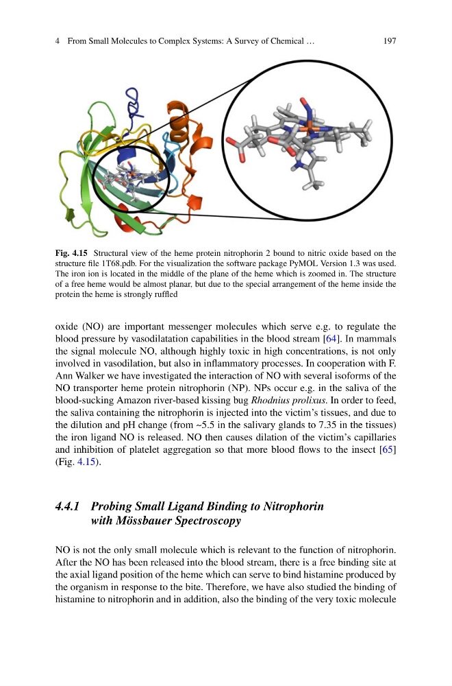
Fig. 4.15 Structural view of the heme protein nitrophorin 2 bound to nitric oxide based on the
structure file 1T68.pdb. For the visualization the software package PyMOL Version 1.3 was used.
The iron ion is located in the middle of the plane of the heme which is zoomed in. The structure
of a free heme would be almost planar, but due to the special arrangement of the heme inside the
protein the heme is strongly ruffled
oxide (NO) are important messenger molecules which serve e.g. to regulate the
blood pressure by vasodilatation capabilities in the blood stream [64]. In mammals
the signal molecule NO, although highly toxic in high concentrations, is not only
involved in vasodilation, but also in inflammatory processes. In cooperation with F.
Ann Walker we have investigated the interaction of NO with several isoforms of the
NO transporter heme protein nitrophorin (NP). NPs occur e.g. in the saliva of the
blood-sucking Amazon river-based kissing bug Rhodnius prolixus. In order to feed,
the saliva containing the nitrophorin is injected into the victim’s tissues, and due to
the dilution and pH change (from ~5.5 in the salivary glands to 7.35 in the tissues)
the iron ligand NO is released. NO then causes dilation of the victim’s capillaries
and inhibition of platelet aggregation so that more blood flows to the insect [65]
(Fig. 4.15).
4.4.1 Probing Small Ligand Binding to Nitrophorin
with Mössbauer Spectroscopy
NO is not the only small molecule which is relevant to the function of nitrophorin.
After the NO has been released into the blood stream, there is a free binding site at
the axial ligand position of the heme which can serve to bind histamine produced by
the organism in response to the bite. Therefore, we have also studied the binding of
histamine to nitrophorin and in addition, also the binding of the very toxic molecule
197
Fig. 4.15 Structural view of the heme protein nitrophorin 2 bound to nitric oxide based on the
structure file 1T68.pdb. For the visualization the software package PyMOL Version 1.3 was used.
The iron ion is located in the middle of the plane of the heme which is zoomed in. The structure
of a free heme would be almost planar, but due to the special arrangement of the heme inside the
protein the heme is strongly ruffled
oxide (NO) are important messenger molecules which serve e.g. to regulate the
blood pressure by vasodilatation capabilities in the blood stream [64]. In mammals
the signal molecule NO, although highly toxic in high concentrations, is not only
involved in vasodilation, but also in inflammatory processes. In cooperation with F.
Ann Walker we have investigated the interaction of NO with several isoforms of the
NO transporter heme protein nitrophorin (NP). NPs occur e.g. in the saliva of the
blood-sucking Amazon river-based kissing bug Rhodnius prolixus. In order to feed,
the saliva containing the nitrophorin is injected into the victim’s tissues, and due to
the dilution and pH change (from ~5.5 in the salivary glands to 7.35 in the tissues)
the iron ligand NO is released. NO then causes dilation of the victim’s capillaries
and inhibition of platelet aggregation so that more blood flows to the insect [65]
(Fig. 4.15).
4.4.1 Probing Small Ligand Binding to Nitrophorin
with Mössbauer Spectroscopy
NO is not the only small molecule which is relevant to the function of nitrophorin.
After the NO has been released into the blood stream, there is a free binding site at
the axial ligand position of the heme which can serve to bind histamine produced by
the organism in response to the bite. Therefore, we have also studied the binding of
histamine to nitrophorin and in addition, also the binding of the very toxic molecule