

9 Mössbauer Spectroscopic Studies on Atomic Diffusion in Materials
501
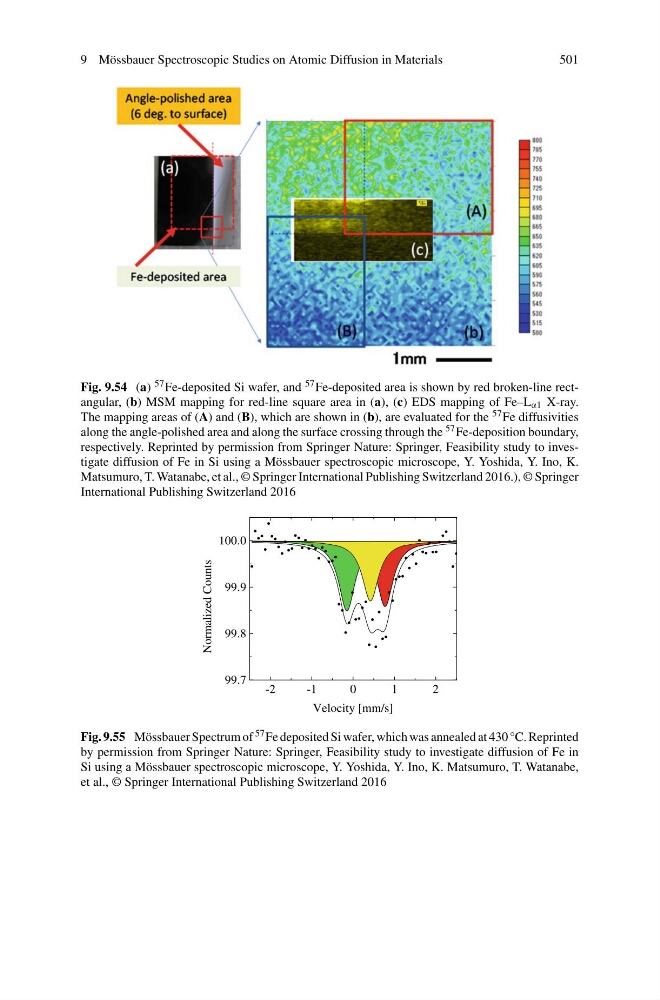
Fig. 9.54 (a) 57 Fe-deposited Si wafer, and 57 Fe-deposited area is shown by red broken-line rectangular, (b) MSM mapping for red-line square area in (a), (c) EDS mapping of Fe–L α1 X-ray.
The mapping areas of (A) and (B), which are shown in (b), are evaluated for the 57 Fe diffusivities
along the angle-polished area and along the surface crossing through the 57 Fe-deposition boundary,
respectively. Reprinted by permission from Springer Nature: Springer, Feasibility study to investigate diffusion of Fe in Si using a Mössbauer spectroscopic microscope, Y. Yoshida, Y. Ino, K.
Matsumuro, T. Watanabe, et al., © Springer International Publishing Switzerland 2016.), © Springer
International Publishing Switzerland 2016
-2
-1
0
1
2
99.7
99.8
99.9
100.0
Normalized Counts
Velocity [mm/s]
Fig. 9.55 Mössbauer Spectrum of 57 Fe deposited Si wafer, which was annealed at 430 °C. Reprinted
by permission from Springer Nature: Springer, Feasibility study to investigate diffusion of Fe in
Si using a Mössbauer spectroscopic microscope, Y. Yoshida, Y. Ino, K. Matsumuro, T. Watanabe,
et al., © Springer International Publishing Switzerland 2016
501
Fig. 9.54 (a) 57 Fe-deposited Si wafer, and 57 Fe-deposited area is shown by red broken-line rectangular, (b) MSM mapping for red-line square area in (a), (c) EDS mapping of Fe–L α1 X-ray.
The mapping areas of (A) and (B), which are shown in (b), are evaluated for the 57 Fe diffusivities
along the angle-polished area and along the surface crossing through the 57 Fe-deposition boundary,
respectively. Reprinted by permission from Springer Nature: Springer, Feasibility study to investigate diffusion of Fe in Si using a Mössbauer spectroscopic microscope, Y. Yoshida, Y. Ino, K.
Matsumuro, T. Watanabe, et al., © Springer International Publishing Switzerland 2016.), © Springer
International Publishing Switzerland 2016
-2
-1
0
1
2
99.7
99.8
99.9
100.0
Normalized Counts
Velocity [mm/s]
Fig. 9.55 Mössbauer Spectrum of 57 Fe deposited Si wafer, which was annealed at 430 °C. Reprinted
by permission from Springer Nature: Springer, Feasibility study to investigate diffusion of Fe in
Si using a Mössbauer spectroscopic microscope, Y. Yoshida, Y. Ino, K. Matsumuro, T. Watanabe,
et al., © Springer International Publishing Switzerland 2016