

9 Mössbauer Spectroscopic Studies on Atomic Diffusion in Materials
499
9.5.4 Imaging Technique Using sCMOS-Camera
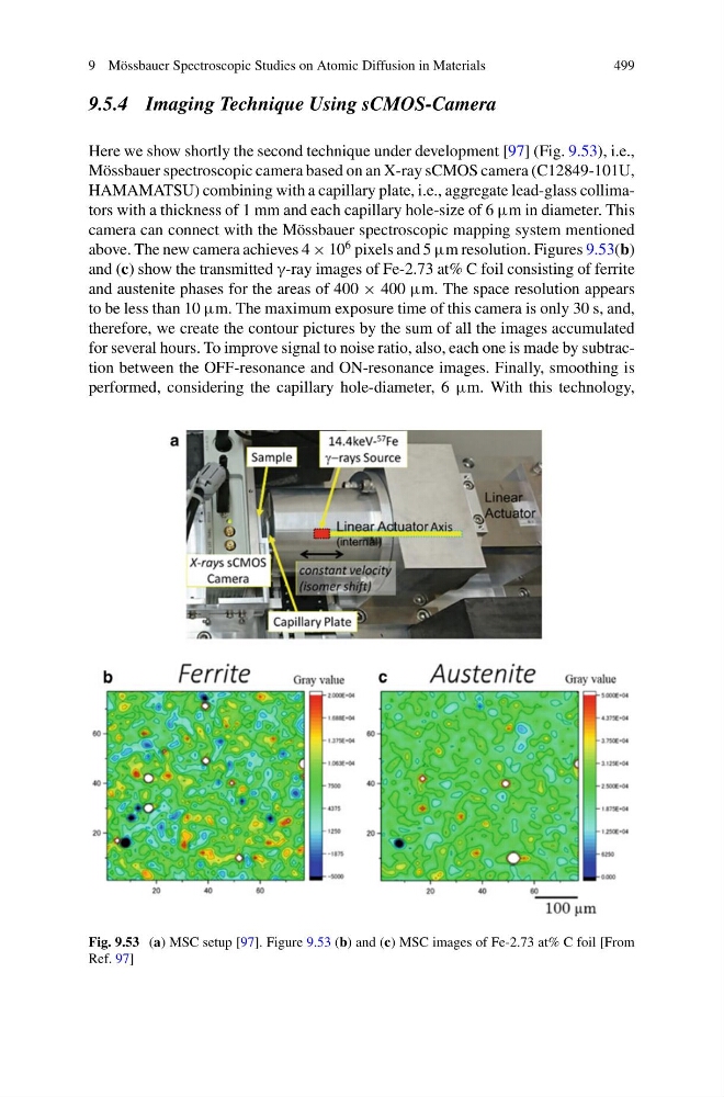
Here we show shortly the second technique under development [97] (Fig. 9.53), i.e.,
Mössbauer spectroscopic camera based on an X-ray sCMOS camera (C12849-101U,
HAMAMATSU) combining with a capillary plate, i.e., aggregate lead-glass collimators with a thickness of 1 mm and each capillary hole-size of 6 μm in diameter. This
camera can connect with the Mössbauer spectroscopic mapping system mentioned
above. The new camera achieves 4 × 10
6 pixels and 5 μm resolution. Figures 9.53(b)
and (c) show the transmitted γ-ray images of Fe-2.73 at% C foil consisting of ferrite
and austenite phases for the areas of 400 × 400 μm. The space resolution appears
to be less than 10 μm. The maximum exposure time of this camera is only 30 s, and,
therefore, we create the contour pictures by the sum of all the images accumulated
for several hours. To improve signal to noise ratio, also, each one is made by subtraction between the OFF-resonance and ON-resonance images. Finally, smoothing is
performed, considering the capillary hole-diameter, 6 μm. With this technology,
Fig. 9.53 (a) MSC setup [97]. Figure 9.53 (b) and (c) MSC images of Fe-2.73 at% C foil [From
Ref. 97]
499
9.5.4 Imaging Technique Using sCMOS-Camera
Here we show shortly the second technique under development [97] (Fig. 9.53), i.e.,
Mössbauer spectroscopic camera based on an X-ray sCMOS camera (C12849-101U,
HAMAMATSU) combining with a capillary plate, i.e., aggregate lead-glass collimators with a thickness of 1 mm and each capillary hole-size of 6 μm in diameter. This
camera can connect with the Mössbauer spectroscopic mapping system mentioned
above. The new camera achieves 4 × 10
6 pixels and 5 μm resolution. Figures 9.53(b)
and (c) show the transmitted γ-ray images of Fe-2.73 at% C foil consisting of ferrite
and austenite phases for the areas of 400 × 400 μm. The space resolution appears
to be less than 10 μm. The maximum exposure time of this camera is only 30 s, and,
therefore, we create the contour pictures by the sum of all the images accumulated
for several hours. To improve signal to noise ratio, also, each one is made by subtraction between the OFF-resonance and ON-resonance images. Finally, smoothing is
performed, considering the capillary hole-diameter, 6 μm. With this technology,
Fig. 9.53 (a) MSC setup [97]. Figure 9.53 (b) and (c) MSC images of Fe-2.73 at% C foil [From
Ref. 97]