

5 Mössbauer Spectroscopy with High Spatial Resolution …
231
5.3.1 Energy and Time Domain Comparison: Spectral
Deconvolution
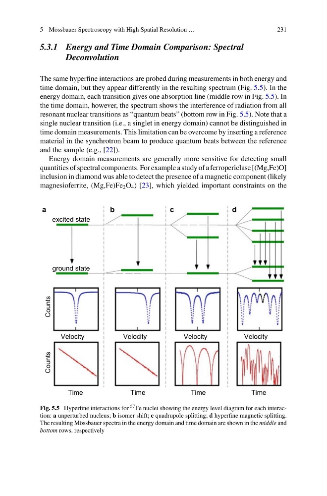
The same hyperfine interactions are probed during measurements in both energy and
time domain, but they appear differently in the resulting spectrum (Fig. 5.5). In the
energy domain, each transition gives one absorption line (middle row in Fig. 5.5). In
the time domain, however, the spectrum shows the interference of radiation from all
resonant nuclear transitions as “quantum beats” (bottom row in Fig. 5.5). Note that a
single nuclear transition (i.e., a singlet in energy domain) cannot be distinguished in
time domain measurements. This limitation can be overcome by inserting a reference
material in the synchrotron beam to produce quantum beats between the reference
and the sample (e.g., [22]).
Energy domain measurements are generally more sensitive for detecting small
quantities of spectral components. For example a study of a ferropericlase [(Mg,Fe)O]
inclusion in diamond was able to detect the presence of a magnetic component (likely
magnesioferrite, (Mg,Fe)Fe 2 O 4 ) [23], which yielded important constraints on the
excited state
ground state
Velocity
Time
Velocity
Time
Velocity
Time
Velocity
Time
Counts
Counts
a
b
c
d
Fig. 5.5 Hyperfine interactions for 57 Fe nuclei showing the energy level diagram for each interaction: a unperturbed nucleus; b isomer shift; c quadrupole splitting; d hyperfine magnetic splitting.
The resulting Mössbauer spectra in the energy domain and time domain are shown in the middle and
bottom rows, respectively
231
5.3.1 Energy and Time Domain Comparison: Spectral
Deconvolution
The same hyperfine interactions are probed during measurements in both energy and
time domain, but they appear differently in the resulting spectrum (Fig. 5.5). In the
energy domain, each transition gives one absorption line (middle row in Fig. 5.5). In
the time domain, however, the spectrum shows the interference of radiation from all
resonant nuclear transitions as “quantum beats” (bottom row in Fig. 5.5). Note that a
single nuclear transition (i.e., a singlet in energy domain) cannot be distinguished in
time domain measurements. This limitation can be overcome by inserting a reference
material in the synchrotron beam to produce quantum beats between the reference
and the sample (e.g., [22]).
Energy domain measurements are generally more sensitive for detecting small
quantities of spectral components. For example a study of a ferropericlase [(Mg,Fe)O]
inclusion in diamond was able to detect the presence of a magnetic component (likely
magnesioferrite, (Mg,Fe)Fe 2 O 4 ) [23], which yielded important constraints on the
excited state
ground state
Velocity
Time
Velocity
Time
Velocity
Time
Velocity
Time
Counts
Counts
a
b
c
d
Fig. 5.5 Hyperfine interactions for 57 Fe nuclei showing the energy level diagram for each interaction: a unperturbed nucleus; b isomer shift; c quadrupole splitting; d hyperfine magnetic splitting.
The resulting Mössbauer spectra in the energy domain and time domain are shown in the middle and
bottom rows, respectively