

3 Quantum Optical Phenomena in Nuclear Resonant Scattering
157
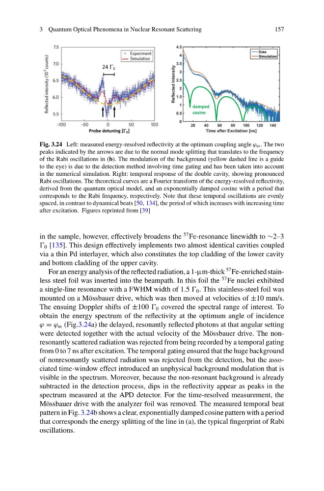
Fig. 3.24 Left: measured energy-resolved reflectivity at the optimum coupling angle ϕ m . The two
peaks indicated by the arrows are due to the normal mode splitting that translates to the frequency
of the Rabi oscillations in (b). The modulation of the background (yellow dashed line is a guide
to the eye) is due to the detection method involving time gating and has been taken into account
in the numerical simulation. Right: temporal response of the double cavity, showing pronounced
Rabi oscillations. The theoretical curves are a Fourier transform of the energy-resolved reflectivity,
derived from the quantum optical model, and an exponentially damped cosine with a period that
corresponds to the Rabi frequency, respectively. Note that these temporal oscillations are evenly
spaced, in contrast to dynamical beats [50, 134], the period of which increases with increasing time
after excitation. Figures reprinted from [39]
in the sample, however, effectively broadens the
57 Fe-resonance linewidth to ∼2–3
0 [135]. This design effectively implements two almost identical cavities coupled
via a thin Pd interlayer, which also constitutes the top cladding of the lower cavity
and bottom cladding of the upper cavity.
For an energy analysis of the reflected radiation, a 1-μm-thick
57 Fe-enriched stainless steel foil was inserted into the beampath. In this foil the
57 Fe nuclei exhibited
a single-line resonance with a FWHM width of 1.5 0 . This stainless-steel foil was
mounted on a Mössbauer drive, which was then moved at velocities of ±10 mm/s.
The ensuing Doppler shifts of ±100 0 covered the spectral range of interest. To
obtain the energy spectrum of the reflectivity at the optimum angle of incidence
ϕ = ϕ m (Fig.3.24a) the delayed, resonantly reflected photons at that angular setting
were detected together with the actual velocity of the Mössbauer drive. The nonresonantly scattered radiation was rejected from being recorded by a temporal gating
from 0 to 7 ns after excitation. The temporal gating ensured that the huge background
of nonresonantly scattered radiation was rejected from the detection, but the associated time-window effect introduced an unphysical background modulation that is
visible in the spectrum. Moreover, because the non-resonant background is already
subtracted in the detection process, dips in the reflectivity appear as peaks in the
spectrum measured at the APD detector. For the time-resolved measurement, the
Mössbauer drive with the analyzer foil was removed. The measured temporal beat
pattern in Fig. 3.24b shows a clear, exponentially damped cosine pattern with a period
that corresponds the energy splitting of the line in (a), the typical fingerprint of Rabi
oscillations.
157
Fig. 3.24 Left: measured energy-resolved reflectivity at the optimum coupling angle ϕ m . The two
peaks indicated by the arrows are due to the normal mode splitting that translates to the frequency
of the Rabi oscillations in (b). The modulation of the background (yellow dashed line is a guide
to the eye) is due to the detection method involving time gating and has been taken into account
in the numerical simulation. Right: temporal response of the double cavity, showing pronounced
Rabi oscillations. The theoretical curves are a Fourier transform of the energy-resolved reflectivity,
derived from the quantum optical model, and an exponentially damped cosine with a period that
corresponds to the Rabi frequency, respectively. Note that these temporal oscillations are evenly
spaced, in contrast to dynamical beats [50, 134], the period of which increases with increasing time
after excitation. Figures reprinted from [39]
in the sample, however, effectively broadens the
57 Fe-resonance linewidth to ∼2–3
0 [135]. This design effectively implements two almost identical cavities coupled
via a thin Pd interlayer, which also constitutes the top cladding of the lower cavity
and bottom cladding of the upper cavity.
For an energy analysis of the reflected radiation, a 1-μm-thick
57 Fe-enriched stainless steel foil was inserted into the beampath. In this foil the
57 Fe nuclei exhibited
a single-line resonance with a FWHM width of 1.5 0 . This stainless-steel foil was
mounted on a Mössbauer drive, which was then moved at velocities of ±10 mm/s.
The ensuing Doppler shifts of ±100 0 covered the spectral range of interest. To
obtain the energy spectrum of the reflectivity at the optimum angle of incidence
ϕ = ϕ m (Fig.3.24a) the delayed, resonantly reflected photons at that angular setting
were detected together with the actual velocity of the Mössbauer drive. The nonresonantly scattered radiation was rejected from being recorded by a temporal gating
from 0 to 7 ns after excitation. The temporal gating ensured that the huge background
of nonresonantly scattered radiation was rejected from the detection, but the associated time-window effect introduced an unphysical background modulation that is
visible in the spectrum. Moreover, because the non-resonant background is already
subtracted in the detection process, dips in the reflectivity appear as peaks in the
spectrum measured at the APD detector. For the time-resolved measurement, the
Mössbauer drive with the analyzer foil was removed. The measured temporal beat
pattern in Fig. 3.24b shows a clear, exponentially damped cosine pattern with a period
that corresponds the energy splitting of the line in (a), the typical fingerprint of Rabi
oscillations.