

neurites, but cRGD still exhibited superior inhibition of neurite expression (Fig. 5).
Therefore, it seems that 2D substrate assessments for DRG growth do not accurately
mimic growth in a 3D environment.
Deister and co-workers investigated neurite extension from explanted dorsal root
ganglia cultured within co-gels made from laminin, fibronectin, collagen I, and HA
[22]. In contrast to previous results, this research showed that apparently neither
collagen nor HA concentration had effects on neurite outgrowth and length. The
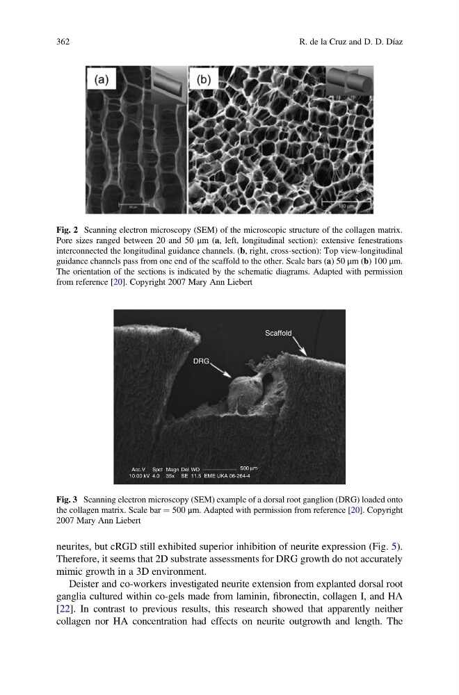
Fig. 2 Scanning electron microscopy (SEM) of the microscopic structure of the collagen matrix.
Pore sizes ranged between 20 and 50 μm (a, left, longitudinal section): extensive fenestrations
interconnected the longitudinal guidance channels. (b, right, cross-section): Top view-longitudinal
guidance channels pass from one end of the scaffold to the other. Scale bars (a) 50 μm (b) 100 μm.
The orientation of the sections is indicated by the schematic diagrams. Adapted with permission
from reference [20]. Copyright 2007 Mary Ann Liebert
Scaffold
DRG
Acc.V Spct Magn
WD
11.5
SE
35x
4.0
10.00 kV
Del
500 µm
EME UKA 06-264-4
Fig. 3 Scanning electron microscopy (SEM) example of a dorsal root ganglion (DRG) loaded onto
the collagen matrix. Scale bar ¼ 500 μm. Adapted with permission from reference [20]. Copyright
2007 Mary Ann Liebert
362
R. de la Cruz and D. D. Díaz
Therefore, it seems that 2D substrate assessments for DRG growth do not accurately
mimic growth in a 3D environment.
Deister and co-workers investigated neurite extension from explanted dorsal root
ganglia cultured within co-gels made from laminin, fibronectin, collagen I, and HA
[22]. In contrast to previous results, this research showed that apparently neither
collagen nor HA concentration had effects on neurite outgrowth and length. The
Fig. 2 Scanning electron microscopy (SEM) of the microscopic structure of the collagen matrix.
Pore sizes ranged between 20 and 50 μm (a, left, longitudinal section): extensive fenestrations
interconnected the longitudinal guidance channels. (b, right, cross-section): Top view-longitudinal
guidance channels pass from one end of the scaffold to the other. Scale bars (a) 50 μm (b) 100 μm.
The orientation of the sections is indicated by the schematic diagrams. Adapted with permission
from reference [20]. Copyright 2007 Mary Ann Liebert
Scaffold
DRG
Acc.V Spct Magn
WD
11.5
SE
35x
4.0
10.00 kV
Del
500 µm
EME UKA 06-264-4
Fig. 3 Scanning electron microscopy (SEM) example of a dorsal root ganglion (DRG) loaded onto
the collagen matrix. Scale bar ¼ 500 μm. Adapted with permission from reference [20]. Copyright
2007 Mary Ann Liebert
362
R. de la Cruz and D. D. Díaz