

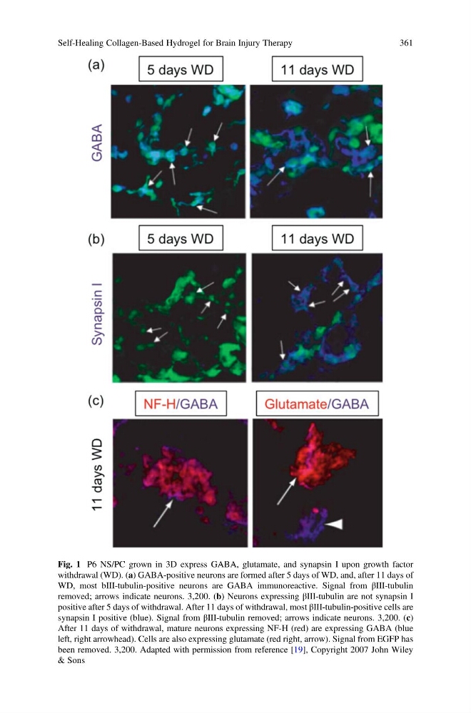
Fig. 1 P6 NS/PC grown in 3D express GABA, glutamate, and synapsin I upon growth factor
withdrawal (WD). (a) GABA-positive neurons are formed after 5 days of WD, and, after 11 days of
WD, most bIII-tubulin-positive neurons are GABA immunoreactive. Signal from βIII-tubulin
removed; arrows indicate neurons. 3,200. (b) Neurons expressing βIII-tubulin are not synapsin I
positive after 5 days of withdrawal. After 11 days of withdrawal, most βIII-tubulin-positive cells are
synapsin I positive (blue). Signal from βIII-tubulin removed; arrows indicate neurons. 3,200. (c)
After 11 days of withdrawal, mature neurons expressing NF-H (red) are expressing GABA (blue
left, right arrowhead). Cells are also expressing glutamate (red right, arrow). Signal from EGFP has
been removed. 3,200. Adapted with permission from reference [19], Copyright 2007 John Wiley
& Sons
Self-Healing Collagen-Based Hydrogel for Brain Injury Therapy
361
withdrawal (WD). (a) GABA-positive neurons are formed after 5 days of WD, and, after 11 days of
WD, most bIII-tubulin-positive neurons are GABA immunoreactive. Signal from βIII-tubulin
removed; arrows indicate neurons. 3,200. (b) Neurons expressing βIII-tubulin are not synapsin I
positive after 5 days of withdrawal. After 11 days of withdrawal, most βIII-tubulin-positive cells are
synapsin I positive (blue). Signal from βIII-tubulin removed; arrows indicate neurons. 3,200. (c)
After 11 days of withdrawal, mature neurons expressing NF-H (red) are expressing GABA (blue
left, right arrowhead). Cells are also expressing glutamate (red right, arrow). Signal from EGFP has
been removed. 3,200. Adapted with permission from reference [19], Copyright 2007 John Wiley
& Sons
Self-Healing Collagen-Based Hydrogel for Brain Injury Therapy
361