

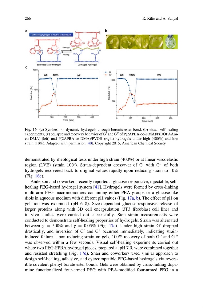
demonstrated by rheological tests under high strain (400%) or at linear viscoelastic
region (LVE) (strain 10%). Strain-dependent crossover of G
0 with G
00 of both
hydrogels recovered back to original values rapidly upon reducing strain to 10%
(Fig. 16c).
Anderson and coworkers recently reported a glucose-responsive, injectable, selfhealing PEG-based hydrogel system [41]. Hydrogels were formed by cross-linking
multi-arm PEG macromonomers containing either PBA groups or a glucose-like
diols in aqueous medium with different pH values (Fig. 17a, b). The effect of pH on
gelation was examined (pH 6–8). Size-dependent glucose-responsive release of
larger proteins along with 3D cell encapsulation (3T3 fibroblast cell line) and
in vivo studies were carried out successfully. Step strain measurements were
conducted to demonstrate self-healing properties of hydrogels. Strain was alternated
between γ ¼ 500% and γ ¼ 0.05% (Fig. 17c). Under high strain G
0 dropped
drastically, and inversion of G
0 and G
00 occurred immediately, indicating straininduced failure. Upon reducing strain on gels, 100% recovery of both G
0 and G
00
was observed within a few seconds. Visual self-healing experiments carried out
where two PEG-FPBA hydrogel pieces, prepared at pH 7.0, were combined together
and resisted stretching (Fig. 17d). Shan and coworkers used similar approach to
design self-healing, adhesive, and cytocompatible PEG-based hydrogels via reversible covalent phenyl borate ester bonds. Gels were obtained by cross-linking dopamine functionalized four-armed PEG with PBA-modified four-armed PEG in a
Fig. 16 (a) Synthesis of dynamic hydrogels through boronic ester bond, (b) visual self-healing
experiments, (c) collapse and recovery behavior of G
0 and G
00 of P(2APBA-co-DMA)/P(DOPAAmco-DMA) (left) and P(2APBA-co-DMA)/PVOH (right) hydrogels under high (400%) and low
strain (10%). Adapted with permission [40]. Copyright 2015, American Chemical Society
266
R. Kilic and A. Sanyal
region (LVE) (strain 10%). Strain-dependent crossover of G
0 with G
00 of both
hydrogels recovered back to original values rapidly upon reducing strain to 10%
(Fig. 16c).
Anderson and coworkers recently reported a glucose-responsive, injectable, selfhealing PEG-based hydrogel system [41]. Hydrogels were formed by cross-linking
multi-arm PEG macromonomers containing either PBA groups or a glucose-like
diols in aqueous medium with different pH values (Fig. 17a, b). The effect of pH on
gelation was examined (pH 6–8). Size-dependent glucose-responsive release of
larger proteins along with 3D cell encapsulation (3T3 fibroblast cell line) and
in vivo studies were carried out successfully. Step strain measurements were
conducted to demonstrate self-healing properties of hydrogels. Strain was alternated
between γ ¼ 500% and γ ¼ 0.05% (Fig. 17c). Under high strain G
0 dropped
drastically, and inversion of G
0 and G
00 occurred immediately, indicating straininduced failure. Upon reducing strain on gels, 100% recovery of both G
0 and G
00
was observed within a few seconds. Visual self-healing experiments carried out
where two PEG-FPBA hydrogel pieces, prepared at pH 7.0, were combined together
and resisted stretching (Fig. 17d). Shan and coworkers used similar approach to
design self-healing, adhesive, and cytocompatible PEG-based hydrogels via reversible covalent phenyl borate ester bonds. Gels were obtained by cross-linking dopamine functionalized four-armed PEG with PBA-modified four-armed PEG in a
Fig. 16 (a) Synthesis of dynamic hydrogels through boronic ester bond, (b) visual self-healing
experiments, (c) collapse and recovery behavior of G
0 and G
00 of P(2APBA-co-DMA)/P(DOPAAmco-DMA) (left) and P(2APBA-co-DMA)/PVOH (right) hydrogels under high (400%) and low
strain (10%). Adapted with permission [40]. Copyright 2015, American Chemical Society
266
R. Kilic and A. Sanyal