

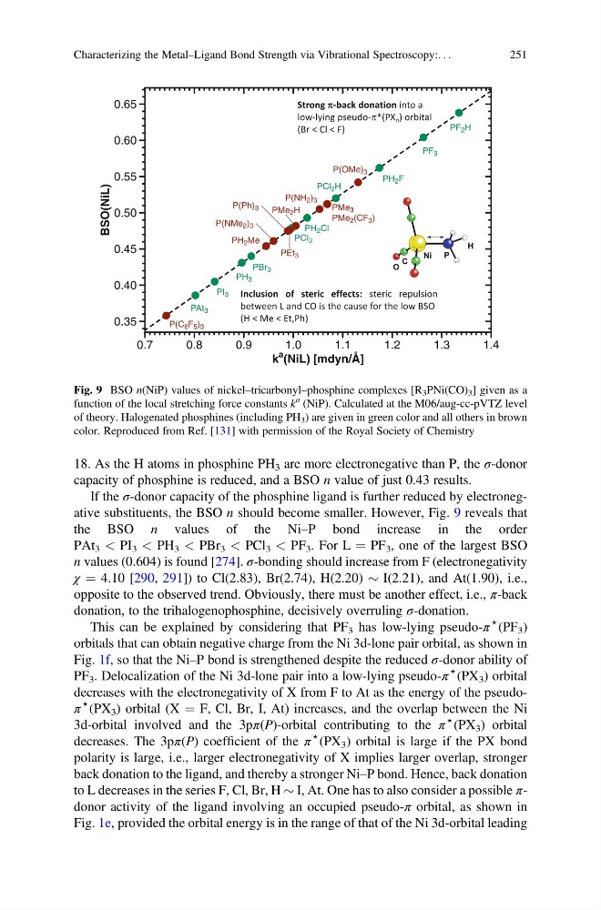
18. As the H atoms in phosphine PH 3 are more electronegative than P, the σ-donor
capacity of phosphine is reduced, and a BSO n value of just 0.43 results.
If the σ-donor capacity of the phosphine ligand is further reduced by electronegative substituents, the BSO n should become smaller. However, Fig. 9 reveals that
the BSO n values of the Ni–P bond increase in the order
PAt 3 < PI 3 < PH 3 < PBr 3 < PCl 3 < PF 3 . For L ¼ PF 3 , one of the largest BSO
n values (0.604) is found [274]. σ-bonding should increase from F (electronegativity
χ ¼ 4.10 [290, 291]) to Cl(2.83), Br(2.74), H(2.20) $ I(2.21), and At(1.90), i.e.,
opposite to the observed trend. Obviously, there must be another effect, i.e., π-back
donation, to the trihalogenophosphine, decisively overruling σ-donation.
This can be explained by considering that PF 3 has low-lying pseudo-π
⋆ (PF 3 )
orbitals that can obtain negative charge from the Ni 3d-lone pair orbital, as shown in
Fig. 1f, so that the Ni–P bond is strengthened despite the reduced σ-donor ability of
PF 3 . Delocalization of the Ni 3d-lone pair into a low-lying pseudo-π
⋆ (PX 3 ) orbital
decreases with the electronegativity of X from F to At as the energy of the pseudoπ
⋆ (PX 3 ) orbital (X ¼ F, Cl, Br, I, At) increases, and the overlap between the Ni
3d-orbital involved and the 3pπ(P)-orbital contributing to the π
⋆ (PX 3 ) orbital
decreases. The 3pπ(P) coefficient of the π
⋆
(PX 3 ) orbital is large if the PX bond
polarity is large, i.e., larger electronegativity of X implies larger overlap, stronger
back donation to the ligand, and thereby a stronger Ni–P bond. Hence, back donation
to L decreases in the series F, Cl, Br, H $ I, At. One has to also consider a possible πdonor activity of the ligand involving an occupied pseudo-π orbital, as shown in
Fig. 1e, provided the orbital energy is in the range of that of the Ni 3d-orbital leading
PF 2 H
PMe 2 H
PEt 3
P(Ph) 3
P(NMe 2 ) 3
PF 3
PH 2 F
PCl 2 H
PBr 3
PH 3
PCl 3
PI 3
PAt 3
P(OMe) 3
PMe 3
PMe 2 (CF 3 )
P(NH 2 ) 3
PH 2 Me
P(C 6 F 5 ) 3
PH 2 Cl
BSO(NiL)
0.35
0.40
0.45
0.50
0.55
0.60
0.65
k
a (NiL) [mdyn/Å]
0.7
0.8
0.9
1.0
1.1
1.2
1.3
1.4
C
O
Ni
P
H
Fig. 9 BSO n(NiP) values of nickel–tricarbonyl–phosphine complexes [R 3 PNi(CO) 3 ] given as a
function of the local stretching force constants k
a (NiP). Calculated at the M06/aug-cc-pVTZ level
of theory. Halogenated phosphines (including PH 3 ) are given in green color and all others in brown
color. Reproduced from Ref. [131] with permission of the Royal Society of Chemistry
Characterizing the Metal–Ligand Bond Strength via Vibrational Spectroscopy:. . .
251
capacity of phosphine is reduced, and a BSO n value of just 0.43 results.
If the σ-donor capacity of the phosphine ligand is further reduced by electronegative substituents, the BSO n should become smaller. However, Fig. 9 reveals that
the BSO n values of the Ni–P bond increase in the order
PAt 3 < PI 3 < PH 3 < PBr 3 < PCl 3 < PF 3 . For L ¼ PF 3 , one of the largest BSO
n values (0.604) is found [274]. σ-bonding should increase from F (electronegativity
χ ¼ 4.10 [290, 291]) to Cl(2.83), Br(2.74), H(2.20) $ I(2.21), and At(1.90), i.e.,
opposite to the observed trend. Obviously, there must be another effect, i.e., π-back
donation, to the trihalogenophosphine, decisively overruling σ-donation.
This can be explained by considering that PF 3 has low-lying pseudo-π
⋆ (PF 3 )
orbitals that can obtain negative charge from the Ni 3d-lone pair orbital, as shown in
Fig. 1f, so that the Ni–P bond is strengthened despite the reduced σ-donor ability of
PF 3 . Delocalization of the Ni 3d-lone pair into a low-lying pseudo-π
⋆ (PX 3 ) orbital
decreases with the electronegativity of X from F to At as the energy of the pseudoπ
⋆ (PX 3 ) orbital (X ¼ F, Cl, Br, I, At) increases, and the overlap between the Ni
3d-orbital involved and the 3pπ(P)-orbital contributing to the π
⋆ (PX 3 ) orbital
decreases. The 3pπ(P) coefficient of the π
⋆
(PX 3 ) orbital is large if the PX bond
polarity is large, i.e., larger electronegativity of X implies larger overlap, stronger
back donation to the ligand, and thereby a stronger Ni–P bond. Hence, back donation
to L decreases in the series F, Cl, Br, H $ I, At. One has to also consider a possible πdonor activity of the ligand involving an occupied pseudo-π orbital, as shown in
Fig. 1e, provided the orbital energy is in the range of that of the Ni 3d-orbital leading
PF 2 H
PMe 2 H
PEt 3
P(Ph) 3
P(NMe 2 ) 3
PF 3
PH 2 F
PCl 2 H
PBr 3
PH 3
PCl 3
PI 3
PAt 3
P(OMe) 3
PMe 3
PMe 2 (CF 3 )
P(NH 2 ) 3
PH 2 Me
P(C 6 F 5 ) 3
PH 2 Cl
BSO(NiL)
0.35
0.40
0.45
0.50
0.55
0.60
0.65
k
a (NiL) [mdyn/Å]
0.7
0.8
0.9
1.0
1.1
1.2
1.3
1.4
C
O
Ni
P
H
Fig. 9 BSO n(NiP) values of nickel–tricarbonyl–phosphine complexes [R 3 PNi(CO) 3 ] given as a
function of the local stretching force constants k
a (NiP). Calculated at the M06/aug-cc-pVTZ level
of theory. Halogenated phosphines (including PH 3 ) are given in green color and all others in brown
color. Reproduced from Ref. [131] with permission of the Royal Society of Chemistry
Characterizing the Metal–Ligand Bond Strength via Vibrational Spectroscopy:. . .
251