

Current Status of the Development of Blood-Based …
187
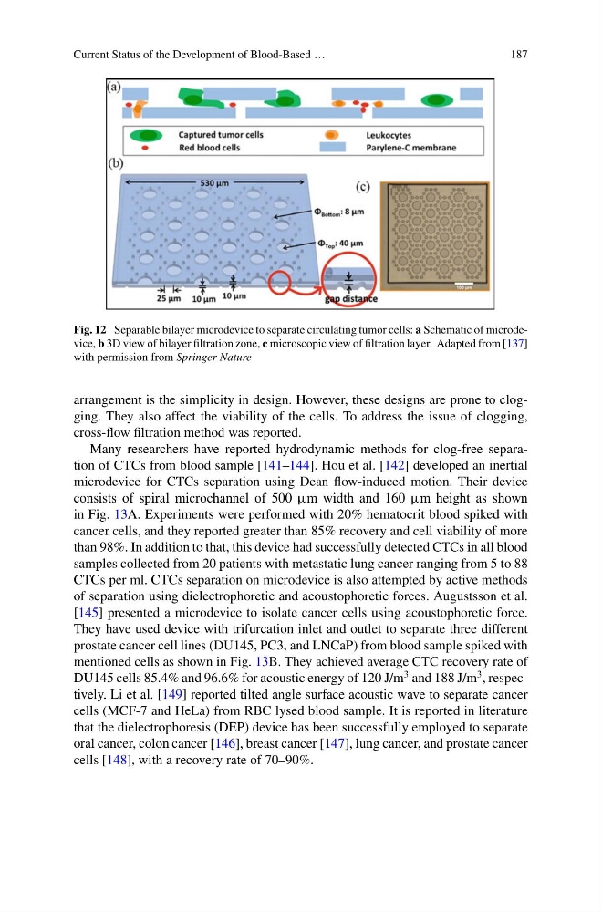
Fig. 12 Separable bilayer microdevice to separate circulating tumor cells: a Schematic of microdevice, b 3D view of bilayer filtration zone, c microscopic view of filtration layer. Adapted from [137]
with permission from Springer Nature
arrangement is the simplicity in design. However, these designs are prone to clogging. They also affect the viability of the cells. To address the issue of clogging,
cross-flow filtration method was reported.
Many researchers have reported hydrodynamic methods for clog-free separation of CTCs from blood sample [141–144]. Hou et al. [142] developed an inertial
microdevice for CTCs separation using Dean flow-induced motion. Their device
consists of spiral microchannel of 500 μm width and 160 μm height as shown
in Fig. 13A. Experiments were performed with 20% hematocrit blood spiked with
cancer cells, and they reported greater than 85% recovery and cell viability of more
than 98%. In addition to that, this device had successfully detected CTCs in all blood
samples collected from 20 patients with metastatic lung cancer ranging from 5 to 88
CTCs per ml. CTCs separation on microdevice is also attempted by active methods
of separation using dielectrophoretic and acoustophoretic forces. Augustsson et al.
[145] presented a microdevice to isolate cancer cells using acoustophoretic force.
They have used device with trifurcation inlet and outlet to separate three different
prostate cancer cell lines (DU145, PC3, and LNCaP) from blood sample spiked with
mentioned cells as shown in Fig. 13B. They achieved average CTC recovery rate of
DU145 cells 85.4% and 96.6% for acoustic energy of 120 J/m
3 and 188 J/m
3 , respectively. Li et al. [149] reported tilted angle surface acoustic wave to separate cancer
cells (MCF-7 and HeLa) from RBC lysed blood sample. It is reported in literature
that the dielectrophoresis (DEP) device has been successfully employed to separate
oral cancer, colon cancer [146], breast cancer [147], lung cancer, and prostate cancer
cells [148], with a recovery rate of 70–90%.
187
Fig. 12 Separable bilayer microdevice to separate circulating tumor cells: a Schematic of microdevice, b 3D view of bilayer filtration zone, c microscopic view of filtration layer. Adapted from [137]
with permission from Springer Nature
arrangement is the simplicity in design. However, these designs are prone to clogging. They also affect the viability of the cells. To address the issue of clogging,
cross-flow filtration method was reported.
Many researchers have reported hydrodynamic methods for clog-free separation of CTCs from blood sample [141–144]. Hou et al. [142] developed an inertial
microdevice for CTCs separation using Dean flow-induced motion. Their device
consists of spiral microchannel of 500 μm width and 160 μm height as shown
in Fig. 13A. Experiments were performed with 20% hematocrit blood spiked with
cancer cells, and they reported greater than 85% recovery and cell viability of more
than 98%. In addition to that, this device had successfully detected CTCs in all blood
samples collected from 20 patients with metastatic lung cancer ranging from 5 to 88
CTCs per ml. CTCs separation on microdevice is also attempted by active methods
of separation using dielectrophoretic and acoustophoretic forces. Augustsson et al.
[145] presented a microdevice to isolate cancer cells using acoustophoretic force.
They have used device with trifurcation inlet and outlet to separate three different
prostate cancer cell lines (DU145, PC3, and LNCaP) from blood sample spiked with
mentioned cells as shown in Fig. 13B. They achieved average CTC recovery rate of
DU145 cells 85.4% and 96.6% for acoustic energy of 120 J/m
3 and 188 J/m
3 , respectively. Li et al. [149] reported tilted angle surface acoustic wave to separate cancer
cells (MCF-7 and HeLa) from RBC lysed blood sample. It is reported in literature
that the dielectrophoresis (DEP) device has been successfully employed to separate
oral cancer, colon cancer [146], breast cancer [147], lung cancer, and prostate cancer
cells [148], with a recovery rate of 70–90%.