

Current Status of the Development of Blood-Based …
185
detect the coagulation time. They used PDMS-based arrangement and applied SAW
to mix citrated blood. Thereafter, image analysis was performed to check the coagulation time. Nam et al. [121] presented Lamb wave-based acoustic streaming of the
particle to measure the coagulation time of blood. They used Lamb wave for instant
mixing of citrated plasma and coagulation reagents in a droplet. After that, acoustic
streaming of florescent particles suspended in plasma was performed to measure the
coagulation time. Coagulation tests were also performed using acoustic resonator
[122].
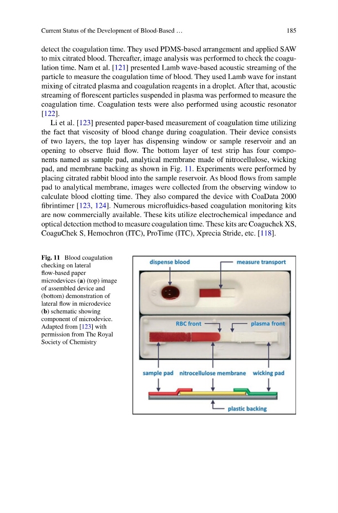
Li et al. [123] presented paper-based measurement of coagulation time utilizing
the fact that viscosity of blood change during coagulation. Their device consists
of two layers, the top layer has dispensing window or sample reservoir and an
opening to observe fluid flow. The bottom layer of test strip has four components named as sample pad, analytical membrane made of nitrocellulose, wicking
pad, and membrane backing as shown in Fig. 11. Experiments were performed by
placing citrated rabbit blood into the sample reservoir. As blood flows from sample
pad to analytical membrane, images were collected from the observing window to
calculate blood clotting time. They also compared the device with CoaData 2000
fibrintimer [123, 124]. Numerous microfluidics-based coagulation monitoring kits
are now commercially available. These kits utilize electrochemical impedance and
optical detection method to measure coagulation time. These kits are Coaguchek XS,
CoaguChek S, Hemochron (ITC), ProTime (ITC), Xprecia Stride, etc. [118].
Fig. 11 Blood coagulation
checking on lateral
flow-based paper
microdevices (a) (top) image
of assembled device and
(bottom) demonstration of
lateral flow in microdevice
(b) schematic showing
component of microdevice.
Adapted from [123] with
permission from The Royal
Society of Chemistry
185
detect the coagulation time. They used PDMS-based arrangement and applied SAW
to mix citrated blood. Thereafter, image analysis was performed to check the coagulation time. Nam et al. [121] presented Lamb wave-based acoustic streaming of the
particle to measure the coagulation time of blood. They used Lamb wave for instant
mixing of citrated plasma and coagulation reagents in a droplet. After that, acoustic
streaming of florescent particles suspended in plasma was performed to measure the
coagulation time. Coagulation tests were also performed using acoustic resonator
[122].
Li et al. [123] presented paper-based measurement of coagulation time utilizing
the fact that viscosity of blood change during coagulation. Their device consists
of two layers, the top layer has dispensing window or sample reservoir and an
opening to observe fluid flow. The bottom layer of test strip has four components named as sample pad, analytical membrane made of nitrocellulose, wicking
pad, and membrane backing as shown in Fig. 11. Experiments were performed by
placing citrated rabbit blood into the sample reservoir. As blood flows from sample
pad to analytical membrane, images were collected from the observing window to
calculate blood clotting time. They also compared the device with CoaData 2000
fibrintimer [123, 124]. Numerous microfluidics-based coagulation monitoring kits
are now commercially available. These kits utilize electrochemical impedance and
optical detection method to measure coagulation time. These kits are Coaguchek XS,
CoaguChek S, Hemochron (ITC), ProTime (ITC), Xprecia Stride, etc. [118].
Fig. 11 Blood coagulation
checking on lateral
flow-based paper
microdevices (a) (top) image
of assembled device and
(bottom) demonstration of
lateral flow in microdevice
(b) schematic showing
component of microdevice.
Adapted from [123] with
permission from The Royal
Society of Chemistry