

184
V. Laxmi et al.
hydrodynamic methods [115], and filtration techniques [116], and affinity-based
method [117]. A brief review of those methods which aims point-of-care analysis is
provided in the following paragraph.
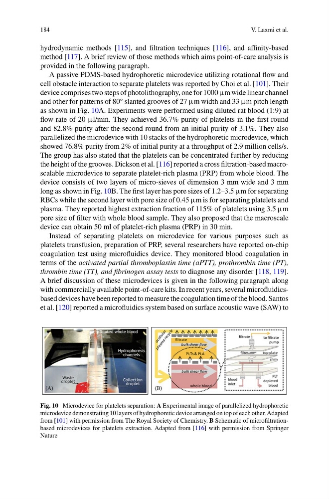
A passive PDMS-based hydrophoretic microdevice utilizing rotational flow and
cell obstacle interaction to separate platelets was reported by Choi et al. [101]. Their
device comprises two steps of photolithography, one for 1000 μm wide linear channel
and other for patterns of 80° slanted grooves of 27 μm width and 33 μm pitch length
as shown in Fig. 10A. Experiments were performed using diluted rat blood (1:9) at
flow rate of 20 μl/min. They achieved 36.7% purity of platelets in the first round
and 82.8% purity after the second round from an initial purity of 3.1%. They also
parallelized the microdevice with 10 stacks of the hydrophoretic microdevice, which
showed 76.8% purity from 2% of initial purity at a throughput of 2.9 million cells/s.
The group has also stated that the platelets can be concentrated further by reducing
the height of the grooves. Dickson et al. [116] reported a cross filtration-based macroscalable microdevice to separate platelet-rich plasma (PRP) from whole blood. The
device consists of two layers of micro-sieves of dimension 3 mm wide and 3 mm
long as shown in Fig. 10B. The first layer has pore sizes of 1.2–3.5 μm for separating
RBCs while the second layer with pore size of 0.45 μm is for separating platelets and
plasma. They reported highest extraction fraction of 115% of platelets using 3.5 μm
pore size of filter with whole blood sample. They also proposed that the macroscale
device can obtain 50 ml of platelet-rich plasma (PRP) in 30 min.
Instead of separating platelets on microdevice for various purposes such as
platelets transfusion, preparation of PRP, several researchers have reported on-chip
coagulation test using microfluidics device. They monitored blood coagulation in
terms of the activated partial thromboplastin time (aPTT), prothrombin time (PT),
thrombin time (TT), and fibrinogen assay tests to diagnose any disorder [118, 119].
A brief discussion of these microdevices is given in the following paragraph along
with commercially available point-of-care kits. In recent years, several microfluidicsbased devices have been reported to measure the coagulation time of the blood. Santos
et al. [120] reported a microfluidics system based on surface acoustic wave (SAW) to
Fig. 10 Microdevice for platelets separation: A Experimental image of parallelized hydrophoretic
microdevice demonstrating 10 layers of hydrophoretic device arranged on top of each other. Adapted
from [101] with permission from The Royal Society of Chemistry. B Schematic of microfiltrationbased microdevices for platelets extraction. Adapted from [116] with permission from Springer
Nature
V. Laxmi et al.
hydrodynamic methods [115], and filtration techniques [116], and affinity-based
method [117]. A brief review of those methods which aims point-of-care analysis is
provided in the following paragraph.
A passive PDMS-based hydrophoretic microdevice utilizing rotational flow and
cell obstacle interaction to separate platelets was reported by Choi et al. [101]. Their
device comprises two steps of photolithography, one for 1000 μm wide linear channel
and other for patterns of 80° slanted grooves of 27 μm width and 33 μm pitch length
as shown in Fig. 10A. Experiments were performed using diluted rat blood (1:9) at
flow rate of 20 μl/min. They achieved 36.7% purity of platelets in the first round
and 82.8% purity after the second round from an initial purity of 3.1%. They also
parallelized the microdevice with 10 stacks of the hydrophoretic microdevice, which
showed 76.8% purity from 2% of initial purity at a throughput of 2.9 million cells/s.
The group has also stated that the platelets can be concentrated further by reducing
the height of the grooves. Dickson et al. [116] reported a cross filtration-based macroscalable microdevice to separate platelet-rich plasma (PRP) from whole blood. The
device consists of two layers of micro-sieves of dimension 3 mm wide and 3 mm
long as shown in Fig. 10B. The first layer has pore sizes of 1.2–3.5 μm for separating
RBCs while the second layer with pore size of 0.45 μm is for separating platelets and
plasma. They reported highest extraction fraction of 115% of platelets using 3.5 μm
pore size of filter with whole blood sample. They also proposed that the macroscale
device can obtain 50 ml of platelet-rich plasma (PRP) in 30 min.
Instead of separating platelets on microdevice for various purposes such as
platelets transfusion, preparation of PRP, several researchers have reported on-chip
coagulation test using microfluidics device. They monitored blood coagulation in
terms of the activated partial thromboplastin time (aPTT), prothrombin time (PT),
thrombin time (TT), and fibrinogen assay tests to diagnose any disorder [118, 119].
A brief discussion of these microdevices is given in the following paragraph along
with commercially available point-of-care kits. In recent years, several microfluidicsbased devices have been reported to measure the coagulation time of the blood. Santos
et al. [120] reported a microfluidics system based on surface acoustic wave (SAW) to
Fig. 10 Microdevice for platelets separation: A Experimental image of parallelized hydrophoretic
microdevice demonstrating 10 layers of hydrophoretic device arranged on top of each other. Adapted
from [101] with permission from The Royal Society of Chemistry. B Schematic of microfiltrationbased microdevices for platelets extraction. Adapted from [116] with permission from Springer
Nature