

Current Status of the Development of Blood-Based …
175
bration curves were generated by measuring the color intensity and intensity versus
concentration graphs were generated for each analyte (AST, ALP, and protein).
Researchers claim that using these calibration curves color intensity charts can
be prepared for easy readouts of the liver tests. This device is low in cost and easy to
operate in resource poor setting, and it also satisfies the ASSURED criteria developed
by WHO.
Tripathi et al. [7] developed a hydrodynamic-based microfluidic device utilizing
biophysical and geometrical effects for plasma extraction. They utilized combination of Fahraeus effect [34–36], centrifugal effect, bifurcation law, and constriction–
expansion zone to separate high purity plasma for wide range of hematocrits. As
shown in Fig. 4B, as the blood flows through a bend the centrifugal forces push
the cells toward the outer wall of the channel and subsequently presence of an
expansion region enhances the cell-free layer. Cell-free plasma flows into the plasma
line after the bifurcation. The separated plasma was subsequently tested (off-chip)
to demonstrate the device capability to detect presence of glucose, hcG hormone
(human chorionic gonadotropin, a pregnancy indicator), and proteins. The detection
of the biomarkers agreed well with the commercially available test strips. Recently,
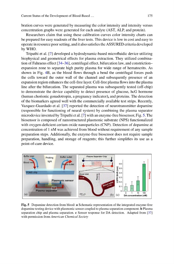
Vazquez-Guardado et al. [37] reported the detection of neurotransmitter dopamine
(responsible for functioning of neural system) by combining the plasma separator
microdevice invented by Tripathi et al. [7] with an enzyme-free biosensor, Fig. 5. The
biosensor is composed of nanostructured plasmonic substrate (NPS) functionalized
with oxygen-deficient cerium oxide nanoparticles (CNP). Detection of dopamine at
concentration of 1 nM was achieved from blood without requirement of any sample
preparation steps. Additionally, the enzyme-free biosensor does not require sample
preparation, handling, and storage of reagents; this further simplifies its use as a
point-of-care device.
Fig. 5 Dopamine detection from blood: a Schematic representation of the integrated enzyme-free
dopamine testing device with plasmonic sensor coupled to plasma separation component. b Plasma
separation chip and plasma separation. c Sensor response for DA detection. Adapted from [37]
with permission from American Chemical Society
175
bration curves were generated by measuring the color intensity and intensity versus
concentration graphs were generated for each analyte (AST, ALP, and protein).
Researchers claim that using these calibration curves color intensity charts can
be prepared for easy readouts of the liver tests. This device is low in cost and easy to
operate in resource poor setting, and it also satisfies the ASSURED criteria developed
by WHO.
Tripathi et al. [7] developed a hydrodynamic-based microfluidic device utilizing
biophysical and geometrical effects for plasma extraction. They utilized combination of Fahraeus effect [34–36], centrifugal effect, bifurcation law, and constriction–
expansion zone to separate high purity plasma for wide range of hematocrits. As
shown in Fig. 4B, as the blood flows through a bend the centrifugal forces push
the cells toward the outer wall of the channel and subsequently presence of an
expansion region enhances the cell-free layer. Cell-free plasma flows into the plasma
line after the bifurcation. The separated plasma was subsequently tested (off-chip)
to demonstrate the device capability to detect presence of glucose, hcG hormone
(human chorionic gonadotropin, a pregnancy indicator), and proteins. The detection
of the biomarkers agreed well with the commercially available test strips. Recently,
Vazquez-Guardado et al. [37] reported the detection of neurotransmitter dopamine
(responsible for functioning of neural system) by combining the plasma separator
microdevice invented by Tripathi et al. [7] with an enzyme-free biosensor, Fig. 5. The
biosensor is composed of nanostructured plasmonic substrate (NPS) functionalized
with oxygen-deficient cerium oxide nanoparticles (CNP). Detection of dopamine at
concentration of 1 nM was achieved from blood without requirement of any sample
preparation steps. Additionally, the enzyme-free biosensor does not require sample
preparation, handling, and storage of reagents; this further simplifies its use as a
point-of-care device.
Fig. 5 Dopamine detection from blood: a Schematic representation of the integrated enzyme-free
dopamine testing device with plasmonic sensor coupled to plasma separation component. b Plasma
separation chip and plasma separation. c Sensor response for DA detection. Adapted from [37]
with permission from American Chemical Society