

Current Status of the Development of Blood-Based …
173
In the recent past, microfluidic technology has rapidly progressed toward the
development of lab-on-chip technologies and has contributed to point-of-care
microdevices for blood plasma separation. Microfluidic methods for blood plasma
separation can be broadly classified as active and passive methods. Active methods of
plasma separation depend on external forces, these forces could be electrical [11–13],
magnetic [14], acoustic [15, 16], optical, or gravity [17, 18]. The passive separation
methods do not depend on external forces and rely on flow properties and geometry
of the device. The passive methods can be further classified as filtration [19–23] sedimentation [24–27], deterministic lateral displacement [28, 29], and hydrodynamic
methods [7, 30–32]. The major advantages offered by the passive hydrodynamic flow
separation methods are simplicity of design, ease of fabrication, continuous operation, use of relatively high flow rates, and ease of integration with a biosensor [6, 10].
In this section, we discuss microfluidic devices which have been developed exclusively for point-of-care use. We also report devices, which can be used for plasma
separation and analyte detection along with already available point-of-care devices
in the market.
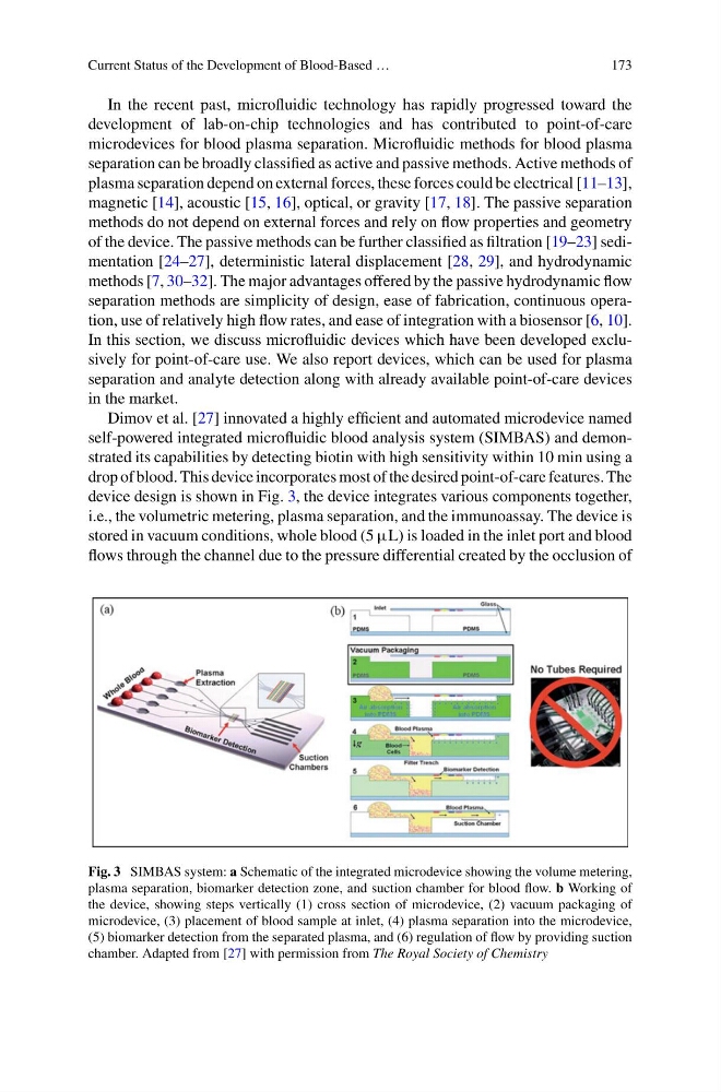
Dimov et al. [27] innovated a highly efficient and automated microdevice named
self-powered integrated microfluidic blood analysis system (SIMBAS) and demonstrated its capabilities by detecting biotin with high sensitivity within 10 min using a
drop of blood. This device incorporates most of the desired point-of-care features. The
device design is shown in Fig. 3, the device integrates various components together,
i.e., the volumetric metering, plasma separation, and the immunoassay. The device is
stored in vacuum conditions, whole blood (5 μL) is loaded in the inlet port and blood
flows through the channel due to the pressure differential created by the occlusion of
Fig. 3 SIMBAS system: a Schematic of the integrated microdevice showing the volume metering,
plasma separation, biomarker detection zone, and suction chamber for blood flow. b Working of
the device, showing steps vertically (1) cross section of microdevice, (2) vacuum packaging of
microdevice, (3) placement of blood sample at inlet, (4) plasma separation into the microdevice,
(5) biomarker detection from the separated plasma, and (6) regulation of flow by providing suction
chamber. Adapted from [27] with permission from The Royal Society of Chemistry
173
In the recent past, microfluidic technology has rapidly progressed toward the
development of lab-on-chip technologies and has contributed to point-of-care
microdevices for blood plasma separation. Microfluidic methods for blood plasma
separation can be broadly classified as active and passive methods. Active methods of
plasma separation depend on external forces, these forces could be electrical [11–13],
magnetic [14], acoustic [15, 16], optical, or gravity [17, 18]. The passive separation
methods do not depend on external forces and rely on flow properties and geometry
of the device. The passive methods can be further classified as filtration [19–23] sedimentation [24–27], deterministic lateral displacement [28, 29], and hydrodynamic
methods [7, 30–32]. The major advantages offered by the passive hydrodynamic flow
separation methods are simplicity of design, ease of fabrication, continuous operation, use of relatively high flow rates, and ease of integration with a biosensor [6, 10].
In this section, we discuss microfluidic devices which have been developed exclusively for point-of-care use. We also report devices, which can be used for plasma
separation and analyte detection along with already available point-of-care devices
in the market.
Dimov et al. [27] innovated a highly efficient and automated microdevice named
self-powered integrated microfluidic blood analysis system (SIMBAS) and demonstrated its capabilities by detecting biotin with high sensitivity within 10 min using a
drop of blood. This device incorporates most of the desired point-of-care features. The
device design is shown in Fig. 3, the device integrates various components together,
i.e., the volumetric metering, plasma separation, and the immunoassay. The device is
stored in vacuum conditions, whole blood (5 μL) is loaded in the inlet port and blood
flows through the channel due to the pressure differential created by the occlusion of
Fig. 3 SIMBAS system: a Schematic of the integrated microdevice showing the volume metering,
plasma separation, biomarker detection zone, and suction chamber for blood flow. b Working of
the device, showing steps vertically (1) cross section of microdevice, (2) vacuum packaging of
microdevice, (3) placement of blood sample at inlet, (4) plasma separation into the microdevice,
(5) biomarker detection from the separated plasma, and (6) regulation of flow by providing suction
chamber. Adapted from [27] with permission from The Royal Society of Chemistry