

594
R. B. Pachori and V. Gupta
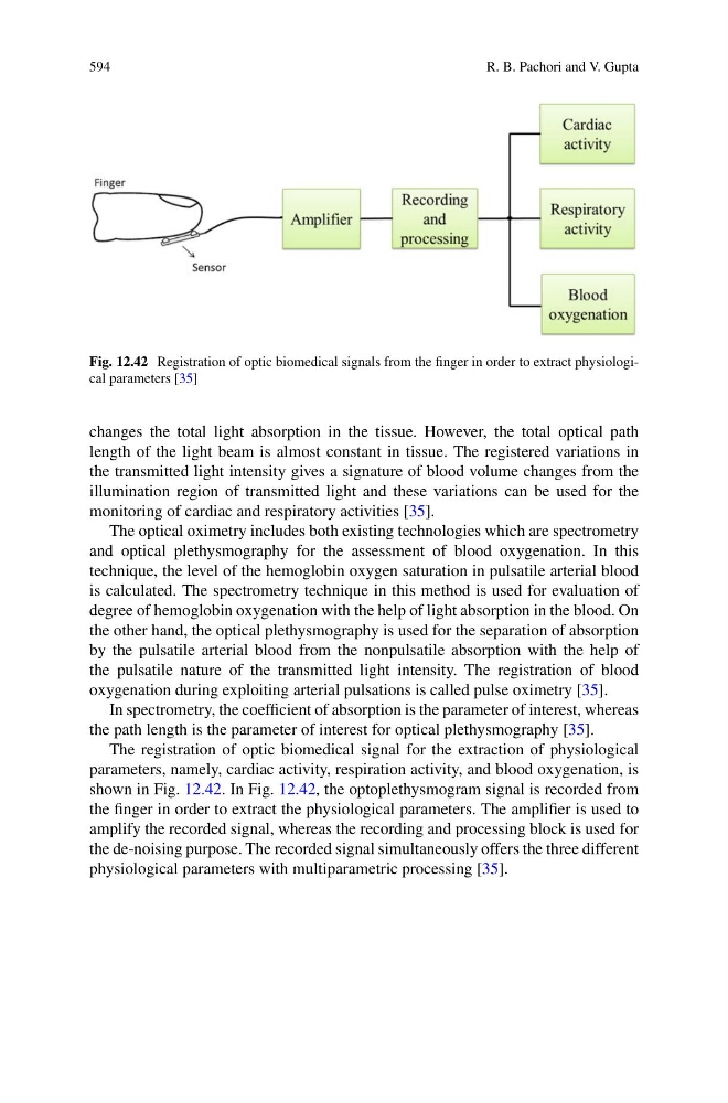
Fig. 12.42 Registration of optic biomedical signals from the finger in order to extract physiological parameters [35]
changes the total light absorption in the tissue. However, the total optical path
length of the light beam is almost constant in tissue. The registered variations in
the transmitted light intensity gives a signature of blood volume changes from the
illumination region of transmitted light and these variations can be used for the
monitoring of cardiac and respiratory activities [35].
The optical oximetry includes both existing technologies which are spectrometry
and optical plethysmography for the assessment of blood oxygenation. In this
technique, the level of the hemoglobin oxygen saturation in pulsatile arterial blood
is calculated. The spectrometry technique in this method is used for evaluation of
degree of hemoglobin oxygenation with the help of light absorption in the blood. On
the other hand, the optical plethysmography is used for the separation of absorption
by the pulsatile arterial blood from the nonpulsatile absorption with the help of
the pulsatile nature of the transmitted light intensity. The registration of blood
oxygenation during exploiting arterial pulsations is called pulse oximetry [35].
In spectrometry, the coefficient of absorption is the parameter of interest, whereas
the path length is the parameter of interest for optical plethysmography [35].
The registration of optic biomedical signal for the extraction of physiological
parameters, namely, cardiac activity, respiration activity, and blood oxygenation, is
shown in Fig. 12.42. In Fig. 12.42, the optoplethysmogram signal is recorded from
the finger in order to extract the physiological parameters. The amplifier is used to
amplify the recorded signal, whereas the recording and processing block is used for
the de-noising purpose. The recorded signal simultaneously offers the three different
physiological parameters with multiparametric processing [35].
R. B. Pachori and V. Gupta
Fig. 12.42 Registration of optic biomedical signals from the finger in order to extract physiological parameters [35]
changes the total light absorption in the tissue. However, the total optical path
length of the light beam is almost constant in tissue. The registered variations in
the transmitted light intensity gives a signature of blood volume changes from the
illumination region of transmitted light and these variations can be used for the
monitoring of cardiac and respiratory activities [35].
The optical oximetry includes both existing technologies which are spectrometry
and optical plethysmography for the assessment of blood oxygenation. In this
technique, the level of the hemoglobin oxygen saturation in pulsatile arterial blood
is calculated. The spectrometry technique in this method is used for evaluation of
degree of hemoglobin oxygenation with the help of light absorption in the blood. On
the other hand, the optical plethysmography is used for the separation of absorption
by the pulsatile arterial blood from the nonpulsatile absorption with the help of
the pulsatile nature of the transmitted light intensity. The registration of blood
oxygenation during exploiting arterial pulsations is called pulse oximetry [35].
In spectrometry, the coefficient of absorption is the parameter of interest, whereas
the path length is the parameter of interest for optical plethysmography [35].
The registration of optic biomedical signal for the extraction of physiological
parameters, namely, cardiac activity, respiration activity, and blood oxygenation, is
shown in Fig. 12.42. In Fig. 12.42, the optoplethysmogram signal is recorded from
the finger in order to extract the physiological parameters. The amplifier is used to
amplify the recorded signal, whereas the recording and processing block is used for
the de-noising purpose. The recorded signal simultaneously offers the three different
physiological parameters with multiparametric processing [35].