

12 Biomedical Engineering Fundamentals
577
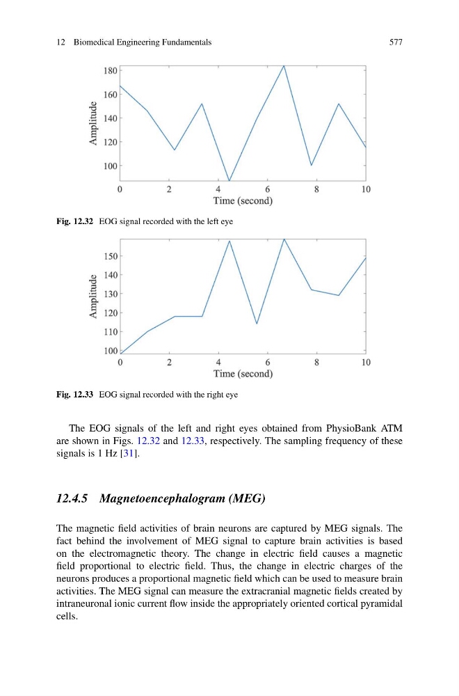
Fig. 12.32 EOG signal recorded with the left eye
Fig. 12.33 EOG signal recorded with the right eye
The EOG signals of the left and right eyes obtained from PhysioBank ATM
are shown in Figs. 12.32 and 12.33, respectively. The sampling frequency of these
signals is 1 Hz [31].
12.4.5 Magnetoencephalogram (MEG)
The magnetic field activities of brain neurons are captured by MEG signals. The
fact behind the involvement of MEG signal to capture brain activities is based
on the electromagnetic theory. The change in electric field causes a magnetic
field proportional to electric field. Thus, the change in electric charges of the
neurons produces a proportional magnetic field which can be used to measure brain
activities. The MEG signal can measure the extracranial magnetic fields created by
intraneuronal ionic current flow inside the appropriately oriented cortical pyramidal
cells.
577
Fig. 12.32 EOG signal recorded with the left eye
Fig. 12.33 EOG signal recorded with the right eye
The EOG signals of the left and right eyes obtained from PhysioBank ATM
are shown in Figs. 12.32 and 12.33, respectively. The sampling frequency of these
signals is 1 Hz [31].
12.4.5 Magnetoencephalogram (MEG)
The magnetic field activities of brain neurons are captured by MEG signals. The
fact behind the involvement of MEG signal to capture brain activities is based
on the electromagnetic theory. The change in electric field causes a magnetic
field proportional to electric field. Thus, the change in electric charges of the
neurons produces a proportional magnetic field which can be used to measure brain
activities. The MEG signal can measure the extracranial magnetic fields created by
intraneuronal ionic current flow inside the appropriately oriented cortical pyramidal
cells.