

307
Characterization and Diagnosis Techniques
combination of ultrahigh voltage beam and high power electron sources
makes the technique destructive for many sample types [17].
In electrochemical supercapacitors, TEM is used to collect and examine the
microstructures of electrode materials, providing information about pore
arrangements [15]. If a specimen is crystalline, the electrons will diffract or
scatter off atomic planes inside the material, enabling the resolution of the
crystal lattice structure down to atomic scale [20]. TEM can also estimate
the level of carbon nanotube (CNT) aggregation or graphene layer thickness.
Similar to SEM, TEM can also show changes in pore structures or arrangements after a material undergoes certain chemical or physical changes
[21,22]. For example, morphological characterization of polymer-dispersed
MWNTs and an image of a carbon template prepared using SBA-15 silica
were characterized using TEM [23].
7.6.3 X-Ray Diffraction (XRD)
XRD is a non-destructive method of bombarding a sample with an x-ray beam
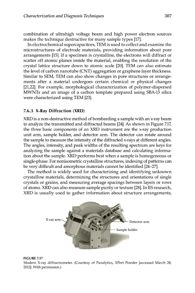
to analyze the transmitted and diffracted beams [24]. As shown in Figure 7.17,
the three basic components of an XRD instrument are the x-ray production
unit arm, sample holder, and detector arm. The detector can rotate around
the sample to measure the intensity of the diffracted x-rays at different angles.
The angles, intensity, and peak widths of the resulting spectrum are keys for
analyzing the sample against a materials database and calculating information about the sample. XRD performs best when a sample is homogeneous or
single-phase. For nonisometric crystalline structures, indexing of patterns can
be very difficult and amorphous materials cannot be identified [24–27].
The method is widely used for characterizing and identifying unknown
crystalline materials, determining the structures and orientations of single
crystals or grains, and measuring average spacings between layers or rows
of atoms. XRD can also measure sample purity or texture [28]. In ES research,
XRD is usually used to gather information about structure arrangements,
X-ray arm
Detector arm
Sample holder
FIGURE 7.17
Modern X-ray diffractrometer. (Courtesy of Panalytics, XPert Powder [accessed March 28,
2012]. With permission.)
Characterization and Diagnosis Techniques
combination of ultrahigh voltage beam and high power electron sources
makes the technique destructive for many sample types [17].
In electrochemical supercapacitors, TEM is used to collect and examine the
microstructures of electrode materials, providing information about pore
arrangements [15]. If a specimen is crystalline, the electrons will diffract or
scatter off atomic planes inside the material, enabling the resolution of the
crystal lattice structure down to atomic scale [20]. TEM can also estimate
the level of carbon nanotube (CNT) aggregation or graphene layer thickness.
Similar to SEM, TEM can also show changes in pore structures or arrangements after a material undergoes certain chemical or physical changes
[21,22]. For example, morphological characterization of polymer-dispersed
MWNTs and an image of a carbon template prepared using SBA-15 silica
were characterized using TEM [23].
7.6.3 X-Ray Diffraction (XRD)
XRD is a non-destructive method of bombarding a sample with an x-ray beam
to analyze the transmitted and diffracted beams [24]. As shown in Figure 7.17,
the three basic components of an XRD instrument are the x-ray production
unit arm, sample holder, and detector arm. The detector can rotate around
the sample to measure the intensity of the diffracted x-rays at different angles.
The angles, intensity, and peak widths of the resulting spectrum are keys for
analyzing the sample against a materials database and calculating information about the sample. XRD performs best when a sample is homogeneous or
single-phase. For nonisometric crystalline structures, indexing of patterns can
be very difficult and amorphous materials cannot be identified [24–27].
The method is widely used for characterizing and identifying unknown
crystalline materials, determining the structures and orientations of single
crystals or grains, and measuring average spacings between layers or rows
of atoms. XRD can also measure sample purity or texture [28]. In ES research,
XRD is usually used to gather information about structure arrangements,
X-ray arm
Detector arm
Sample holder
FIGURE 7.17
Modern X-ray diffractrometer. (Courtesy of Panalytics, XPert Powder [accessed March 28,
2012]. With permission.)