

15 Solid-State Fluorescence Switching Using Photochromic …
317
Δ
Crystal 1a-β
UV
Vis.
Crystal 1a-α
1a
1b
Vis.
Crystal 1b
Crystal 1a′
S
S
F
F
F
F
F
F
Me
Me
S
S
F
F
F
F
F
F
Me
Me
(a)
(b)
(c)
(in crystal)
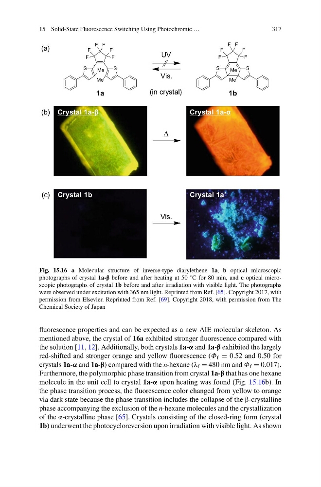
Fig. 15.16 a Molecular structure of inverse-type diarylethene 1a, b optical microscopic
photographs of crystal 1a-β before and after heating at 50 °C for 80 min, and c optical microscopic photographs of crystal 1b before and after irradiation with visible light. The photographs
were observed under excitation with 365 nm light. Reprinted from Ref. [65]. Copyright 2017, with
permission from Elsevier. Reprinted from Ref. [69]. Copyright 2018, with permission from The
Chemical Society of Japan
fluorescence properties and can be expected as a new AIE molecular skeleton. As
mentioned above, the crystal of 16a exhibited stronger fluorescence compared with
the solution [11, 12]. Additionally, both crystals 1a-α and 1a-β exhibited the largely
red-shifted and stronger orange and yellow fluorescence (Φ f = 0.52 and 0.50 for
crystals 1a-α and 1a-β) compared with the n-hexane (λ f = 480 nm and Φ f = 0.017).
Furthermore, the polymorphic phase transition from crystal 1a-β that has one hexane
molecule in the unit cell to crystal 1a-α upon heating was found (Fig. 15.16b). In
the phase transition process, the fluorescence color changed from yellow to orange
via dark state because the phase transition includes the collapse of the β-crystalline
phase accompanying the exclusion of the n-hexane molecules and the crystallization
of the α-crystalline phase [65]. Crystals consisting of the closed-ring form (crystal
1b) underwent the photocycloreversion upon irradiation with visible light. As shown
317
Δ
Crystal 1a-β
UV
Vis.
Crystal 1a-α
1a
1b
Vis.
Crystal 1b
Crystal 1a′
S
S
F
F
F
F
F
F
Me
Me
S
S
F
F
F
F
F
F
Me
Me
(a)
(b)
(c)
(in crystal)
Fig. 15.16 a Molecular structure of inverse-type diarylethene 1a, b optical microscopic
photographs of crystal 1a-β before and after heating at 50 °C for 80 min, and c optical microscopic photographs of crystal 1b before and after irradiation with visible light. The photographs
were observed under excitation with 365 nm light. Reprinted from Ref. [65]. Copyright 2017, with
permission from Elsevier. Reprinted from Ref. [69]. Copyright 2018, with permission from The
Chemical Society of Japan
fluorescence properties and can be expected as a new AIE molecular skeleton. As
mentioned above, the crystal of 16a exhibited stronger fluorescence compared with
the solution [11, 12]. Additionally, both crystals 1a-α and 1a-β exhibited the largely
red-shifted and stronger orange and yellow fluorescence (Φ f = 0.52 and 0.50 for
crystals 1a-α and 1a-β) compared with the n-hexane (λ f = 480 nm and Φ f = 0.017).
Furthermore, the polymorphic phase transition from crystal 1a-β that has one hexane
molecule in the unit cell to crystal 1a-α upon heating was found (Fig. 15.16b). In
the phase transition process, the fluorescence color changed from yellow to orange
via dark state because the phase transition includes the collapse of the β-crystalline
phase accompanying the exclusion of the n-hexane molecules and the crystallization
of the α-crystalline phase [65]. Crystals consisting of the closed-ring form (crystal
1b) underwent the photocycloreversion upon irradiation with visible light. As shown