

260
T. Onodera et al.
would be emerged at the core–shell interface [31], rather than Ag NPs (λ Ag, LSPR
= ca. 390–400 nm). This would expectedly lead to the enhancement of NLO properties [32], which is due to typically so-called excitation enhancement [23]. The
surface of silica-coated PDA NCFs was further modified chemically by using silanecoupling agent (APTES: 3-aminopropyltriethoxysilane), so that amino-functional
groups introduced on the surface of silica layer could control the adsorption amounts
of Au NPs [33, 34]. After adding Au nano-seed (2–3 nm in size), aqueous solution of
Au ion (Au
3+ ) added was reduced mildly by using reducing agent of formaldehyde (HCHO). Here, Au nano-seed was prepared by mixing an aqueous solution containing a given amount of tetrachloroauric (III) acid (H[AuCl] 4 ), sodium
hydroxide (NaOH), and tertakis(hyroxymethyl)phosphonium chloride (THPC) [35].
On the other hand, aqueous solution of Au
3+ was obtained by the incubating the
mixture of H[AuCl] 4 aq. and potassium carbonate (K 2 CO 3 ) aq. under dark condition at ca. 5 °C for three days [33]. Furthermore, the previously Pt-sputtered filter
substrate was employed, when hybridized PDA NCFs was characterized with SEM
observation without using the conventional Pt-sputtering treatment. One can clearly
observe the deposited Au NPs, because Au NPs are distinguished from Pt NPs on
the filter substrate.
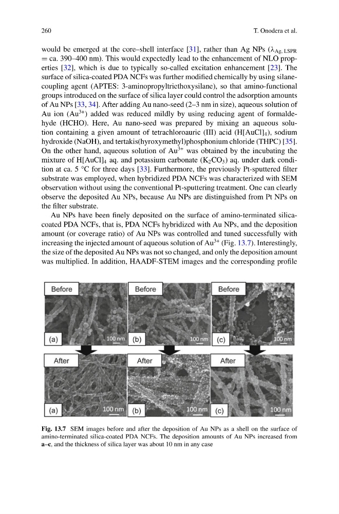
Au NPs have been finely deposited on the surface of amino-terminated silicacoated PDA NCFs, that is, PDA NCFs hybridized with Au NPs, and the deposition
amount (or coverage ratio) of Au NPs was controlled and tuned successfully with
increasing the injected amount of aqueous solution of Au
3+ (Fig. 13.7). Interestingly,
the size of the deposited Au NPs was not so changed, and only the deposition amount
was multiplied. In addition, HAADF-STEM images and the corresponding profile
Fig. 13.7 SEM images before and after the deposition of Au NPs as a shell on the surface of
amino-terminated silica-coated PDA NCFs. The deposition amounts of Au NPs increased from
a–c, and the thickness of silica layer was about 10 nm in any case
T. Onodera et al.
would be emerged at the core–shell interface [31], rather than Ag NPs (λ Ag, LSPR
= ca. 390–400 nm). This would expectedly lead to the enhancement of NLO properties [32], which is due to typically so-called excitation enhancement [23]. The
surface of silica-coated PDA NCFs was further modified chemically by using silanecoupling agent (APTES: 3-aminopropyltriethoxysilane), so that amino-functional
groups introduced on the surface of silica layer could control the adsorption amounts
of Au NPs [33, 34]. After adding Au nano-seed (2–3 nm in size), aqueous solution of
Au ion (Au
3+ ) added was reduced mildly by using reducing agent of formaldehyde (HCHO). Here, Au nano-seed was prepared by mixing an aqueous solution containing a given amount of tetrachloroauric (III) acid (H[AuCl] 4 ), sodium
hydroxide (NaOH), and tertakis(hyroxymethyl)phosphonium chloride (THPC) [35].
On the other hand, aqueous solution of Au
3+ was obtained by the incubating the
mixture of H[AuCl] 4 aq. and potassium carbonate (K 2 CO 3 ) aq. under dark condition at ca. 5 °C for three days [33]. Furthermore, the previously Pt-sputtered filter
substrate was employed, when hybridized PDA NCFs was characterized with SEM
observation without using the conventional Pt-sputtering treatment. One can clearly
observe the deposited Au NPs, because Au NPs are distinguished from Pt NPs on
the filter substrate.
Au NPs have been finely deposited on the surface of amino-terminated silicacoated PDA NCFs, that is, PDA NCFs hybridized with Au NPs, and the deposition
amount (or coverage ratio) of Au NPs was controlled and tuned successfully with
increasing the injected amount of aqueous solution of Au
3+ (Fig. 13.7). Interestingly,
the size of the deposited Au NPs was not so changed, and only the deposition amount
was multiplied. In addition, HAADF-STEM images and the corresponding profile
Fig. 13.7 SEM images before and after the deposition of Au NPs as a shell on the surface of
amino-terminated silica-coated PDA NCFs. The deposition amounts of Au NPs increased from
a–c, and the thickness of silica layer was about 10 nm in any case