

1 X-Ray Birefringence Imaging (XBI): A New Technique …
11
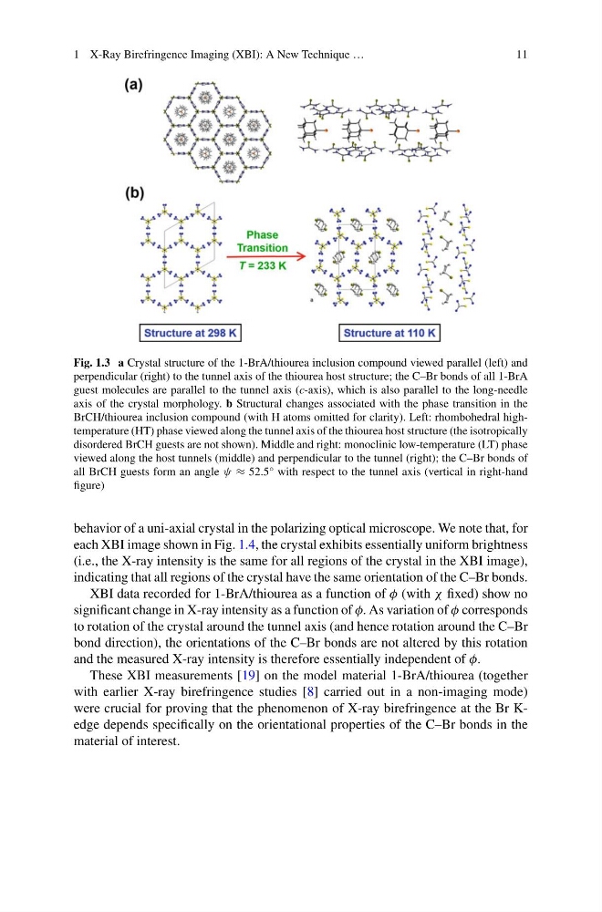
Fig. 1.3 a Crystal structure of the 1-BrA/thiourea inclusion compound viewed parallel (left) and
perpendicular (right) to the tunnel axis of the thiourea host structure; the C–Br bonds of all 1-BrA
guest molecules are parallel to the tunnel axis (c-axis), which is also parallel to the long-needle
axis of the crystal morphology. b Structural changes associated with the phase transition in the
BrCH/thiourea inclusion compound (with H atoms omitted for clarity). Left: rhombohedral hightemperature (HT) phase viewed along the tunnel axis of the thiourea host structure (the isotropically
disordered BrCH guests are not shown). Middle and right: monoclinic low-temperature (LT) phase
viewed along the host tunnels (middle) and perpendicular to the tunnel (right); the C–Br bonds of
all BrCH guests form an angle ψ ≈ 52.5° with respect to the tunnel axis (vertical in right-hand
figure)
behavior of a uni-axial crystal in the polarizing optical microscope. We note that, for
each XBI image shown in Fig. 1.4, the crystal exhibits essentially uniform brightness
(i.e., the X-ray intensity is the same for all regions of the crystal in the XBI image),
indicating that all regions of the crystal have the same orientation of the C–Br bonds.
XBI data recorded for 1-BrA/thiourea as a function of φ (with χ fixed) show no
significant change in X-ray intensity as a function of φ. As variation of φ corresponds
to rotation of the crystal around the tunnel axis (and hence rotation around the C–Br
bond direction), the orientations of the C–Br bonds are not altered by this rotation
and the measured X-ray intensity is therefore essentially independent of φ.
These XBI measurements [19] on the model material 1-BrA/thiourea (together
with earlier X-ray birefringence studies [8] carried out in a non-imaging mode)
were crucial for proving that the phenomenon of X-ray birefringence at the Br Kedge depends specifically on the orientational properties of the C–Br bonds in the
material of interest.
11
Fig. 1.3 a Crystal structure of the 1-BrA/thiourea inclusion compound viewed parallel (left) and
perpendicular (right) to the tunnel axis of the thiourea host structure; the C–Br bonds of all 1-BrA
guest molecules are parallel to the tunnel axis (c-axis), which is also parallel to the long-needle
axis of the crystal morphology. b Structural changes associated with the phase transition in the
BrCH/thiourea inclusion compound (with H atoms omitted for clarity). Left: rhombohedral hightemperature (HT) phase viewed along the tunnel axis of the thiourea host structure (the isotropically
disordered BrCH guests are not shown). Middle and right: monoclinic low-temperature (LT) phase
viewed along the host tunnels (middle) and perpendicular to the tunnel (right); the C–Br bonds of
all BrCH guests form an angle ψ ≈ 52.5° with respect to the tunnel axis (vertical in right-hand
figure)
behavior of a uni-axial crystal in the polarizing optical microscope. We note that, for
each XBI image shown in Fig. 1.4, the crystal exhibits essentially uniform brightness
(i.e., the X-ray intensity is the same for all regions of the crystal in the XBI image),
indicating that all regions of the crystal have the same orientation of the C–Br bonds.
XBI data recorded for 1-BrA/thiourea as a function of φ (with χ fixed) show no
significant change in X-ray intensity as a function of φ. As variation of φ corresponds
to rotation of the crystal around the tunnel axis (and hence rotation around the C–Br
bond direction), the orientations of the C–Br bonds are not altered by this rotation
and the measured X-ray intensity is therefore essentially independent of φ.
These XBI measurements [19] on the model material 1-BrA/thiourea (together
with earlier X-ray birefringence studies [8] carried out in a non-imaging mode)
were crucial for proving that the phenomenon of X-ray birefringence at the Br Kedge depends specifically on the orientational properties of the C–Br bonds in the
material of interest.