

21 Giant Amplification of Fluorescence Quenching in Photochromic …
371
1
2
3
4
5
(i)
(ii)
(iii)
(iv)
(v)
0.0
0.5
1.0
1.5
2.0
2.5
0
50
100
150
200
Intensity / a.u.
Time / sec
UV
UV
438 nm
438 nm
438 nm
(a)
(b)
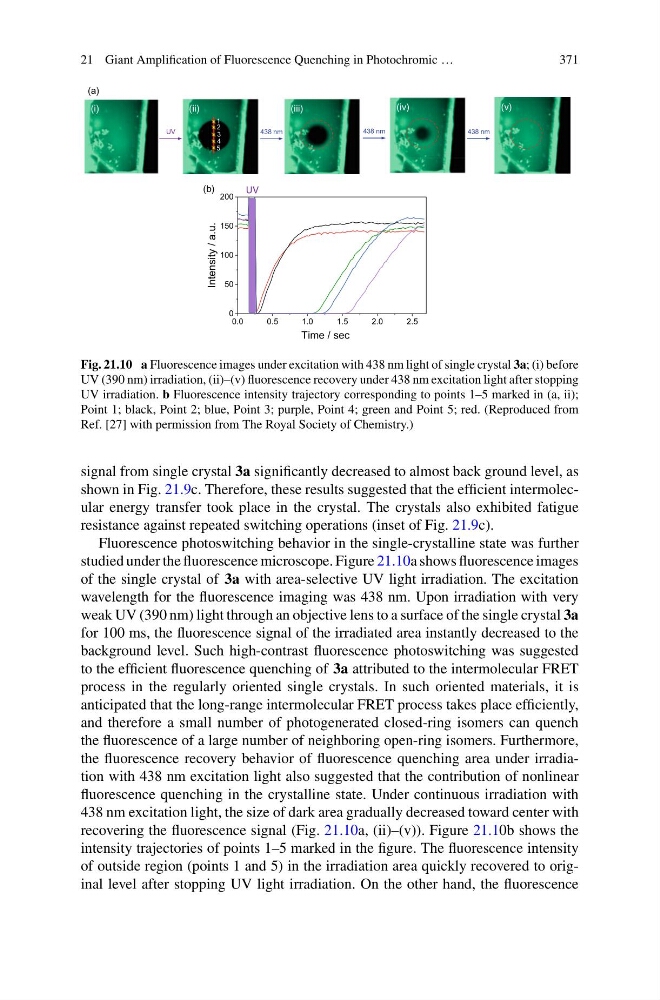
Fig. 21.10 a Fluorescence images under excitation with 438 nm light of single crystal 3a; (i) before
UV (390 nm) irradiation, (ii)–(v) fluorescence recovery under 438 nm excitation light after stopping
UV irradiation. b Fluorescence intensity trajectory corresponding to points 1–5 marked in (a, ii);
Point 1; black, Point 2; blue, Point 3; purple, Point 4; green and Point 5; red. (Reproduced from
Ref. [27] with permission from The Royal Society of Chemistry.)
signal from single crystal 3a significantly decreased to almost back ground level, as
shown in Fig. 21.9c. Therefore, these results suggested that the efficient intermolecular energy transfer took place in the crystal. The crystals also exhibited fatigue
resistance against repeated switching operations (inset of Fig. 21.9c).
Fluorescence photoswitching behavior in the single-crystalline state was further
studied under the fluorescence microscope. Figure 21.10a shows fluorescence images
of the single crystal of 3a with area-selective UV light irradiation. The excitation
wavelength for the fluorescence imaging was 438 nm. Upon irradiation with very
weak UV (390 nm) light through an objective lens to a surface of the single crystal 3a
for 100 ms, the fluorescence signal of the irradiated area instantly decreased to the
background level. Such high-contrast fluorescence photoswitching was suggested
to the efficient fluorescence quenching of 3a attributed to the intermolecular FRET
process in the regularly oriented single crystals. In such oriented materials, it is
anticipated that the long-range intermolecular FRET process takes place efficiently,
and therefore a small number of photogenerated closed-ring isomers can quench
the fluorescence of a large number of neighboring open-ring isomers. Furthermore,
the fluorescence recovery behavior of fluorescence quenching area under irradiation with 438 nm excitation light also suggested that the contribution of nonlinear
fluorescence quenching in the crystalline state. Under continuous irradiation with
438 nm excitation light, the size of dark area gradually decreased toward center with
recovering the fluorescence signal (Fig. 21.10a, (ii)–(v)). Figure 21.10b shows the
intensity trajectories of points 1–5 marked in the figure. The fluorescence intensity
of outside region (points 1 and 5) in the irradiation area quickly recovered to original level after stopping UV light irradiation. On the other hand, the fluorescence
371
1
2
3
4
5
(i)
(ii)
(iii)
(iv)
(v)
0.0
0.5
1.0
1.5
2.0
2.5
0
50
100
150
200
Intensity / a.u.
Time / sec
UV
UV
438 nm
438 nm
438 nm
(a)
(b)
Fig. 21.10 a Fluorescence images under excitation with 438 nm light of single crystal 3a; (i) before
UV (390 nm) irradiation, (ii)–(v) fluorescence recovery under 438 nm excitation light after stopping
UV irradiation. b Fluorescence intensity trajectory corresponding to points 1–5 marked in (a, ii);
Point 1; black, Point 2; blue, Point 3; purple, Point 4; green and Point 5; red. (Reproduced from
Ref. [27] with permission from The Royal Society of Chemistry.)
signal from single crystal 3a significantly decreased to almost back ground level, as
shown in Fig. 21.9c. Therefore, these results suggested that the efficient intermolecular energy transfer took place in the crystal. The crystals also exhibited fatigue
resistance against repeated switching operations (inset of Fig. 21.9c).
Fluorescence photoswitching behavior in the single-crystalline state was further
studied under the fluorescence microscope. Figure 21.10a shows fluorescence images
of the single crystal of 3a with area-selective UV light irradiation. The excitation
wavelength for the fluorescence imaging was 438 nm. Upon irradiation with very
weak UV (390 nm) light through an objective lens to a surface of the single crystal 3a
for 100 ms, the fluorescence signal of the irradiated area instantly decreased to the
background level. Such high-contrast fluorescence photoswitching was suggested
to the efficient fluorescence quenching of 3a attributed to the intermolecular FRET
process in the regularly oriented single crystals. In such oriented materials, it is
anticipated that the long-range intermolecular FRET process takes place efficiently,
and therefore a small number of photogenerated closed-ring isomers can quench
the fluorescence of a large number of neighboring open-ring isomers. Furthermore,
the fluorescence recovery behavior of fluorescence quenching area under irradiation with 438 nm excitation light also suggested that the contribution of nonlinear
fluorescence quenching in the crystalline state. Under continuous irradiation with
438 nm excitation light, the size of dark area gradually decreased toward center with
recovering the fluorescence signal (Fig. 21.10a, (ii)–(v)). Figure 21.10b shows the
intensity trajectories of points 1–5 marked in the figure. The fluorescence intensity
of outside region (points 1 and 5) in the irradiation area quickly recovered to original level after stopping UV light irradiation. On the other hand, the fluorescence