

120
S. Toyouchi et al.
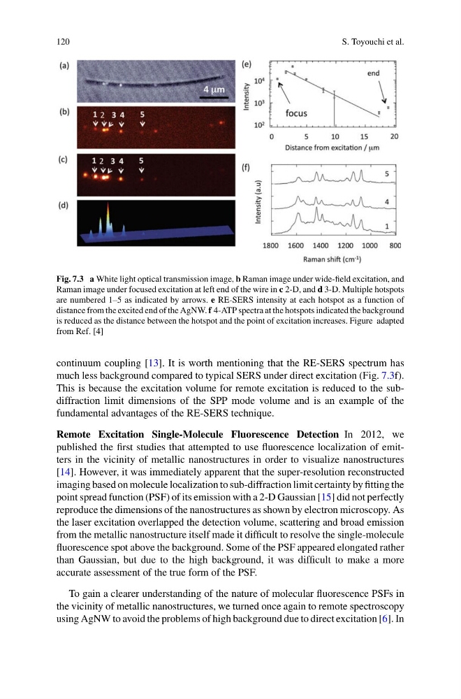
Fig. 7.3 a White light optical transmission image, b Raman image under wide-field excitation, and
Raman image under focused excitation at left end of the wire in c 2-D, and d 3-D. Multiple hotspots
are numbered 1–5 as indicated by arrows. e RE-SERS intensity at each hotspot as a function of
distance from the excited end of the AgNW. f 4-ATP spectra at the hotspots indicated the background
is reduced as the distance between the hotspot and the point of excitation increases. Figure adapted
from Ref. [4]
continuum coupling [13]. It is worth mentioning that the RE-SERS spectrum has
much less background compared to typical SERS under direct excitation (Fig. 7.3f).
This is because the excitation volume for remote excitation is reduced to the subdiffraction limit dimensions of the SPP mode volume and is an example of the
fundamental advantages of the RE-SERS technique.
Remote Excitation Single-Molecule Fluorescence Detection In 2012, we
published the first studies that attempted to use fluorescence localization of emitters in the vicinity of metallic nanostructures in order to visualize nanostructures
[14]. However, it was immediately apparent that the super-resolution reconstructed
imaging based on molecule localization to sub-diffraction limit certainty by fitting the
point spread function (PSF) of its emission with a 2-D Gaussian [15] did not perfectly
reproduce the dimensions of the nanostructures as shown by electron microscopy. As
the laser excitation overlapped the detection volume, scattering and broad emission
from the metallic nanostructure itself made it difficult to resolve the single-molecule
fluorescence spot above the background. Some of the PSF appeared elongated rather
than Gaussian, but due to the high background, it was difficult to make a more
accurate assessment of the true form of the PSF.
To gain a clearer understanding of the nature of molecular fluorescence PSFs in
the vicinity of metallic nanostructures, we turned once again to remote spectroscopy
using AgNW to avoid the problems of high background due to direct excitation [6]. In
S. Toyouchi et al.
Fig. 7.3 a White light optical transmission image, b Raman image under wide-field excitation, and
Raman image under focused excitation at left end of the wire in c 2-D, and d 3-D. Multiple hotspots
are numbered 1–5 as indicated by arrows. e RE-SERS intensity at each hotspot as a function of
distance from the excited end of the AgNW. f 4-ATP spectra at the hotspots indicated the background
is reduced as the distance between the hotspot and the point of excitation increases. Figure adapted
from Ref. [4]
continuum coupling [13]. It is worth mentioning that the RE-SERS spectrum has
much less background compared to typical SERS under direct excitation (Fig. 7.3f).
This is because the excitation volume for remote excitation is reduced to the subdiffraction limit dimensions of the SPP mode volume and is an example of the
fundamental advantages of the RE-SERS technique.
Remote Excitation Single-Molecule Fluorescence Detection In 2012, we
published the first studies that attempted to use fluorescence localization of emitters in the vicinity of metallic nanostructures in order to visualize nanostructures
[14]. However, it was immediately apparent that the super-resolution reconstructed
imaging based on molecule localization to sub-diffraction limit certainty by fitting the
point spread function (PSF) of its emission with a 2-D Gaussian [15] did not perfectly
reproduce the dimensions of the nanostructures as shown by electron microscopy. As
the laser excitation overlapped the detection volume, scattering and broad emission
from the metallic nanostructure itself made it difficult to resolve the single-molecule
fluorescence spot above the background. Some of the PSF appeared elongated rather
than Gaussian, but due to the high background, it was difficult to make a more
accurate assessment of the true form of the PSF.
To gain a clearer understanding of the nature of molecular fluorescence PSFs in
the vicinity of metallic nanostructures, we turned once again to remote spectroscopy
using AgNW to avoid the problems of high background due to direct excitation [6]. In