

86
4 Results and Discussion
PCM microcapsules
on the surface of the foam
The structure of the foam
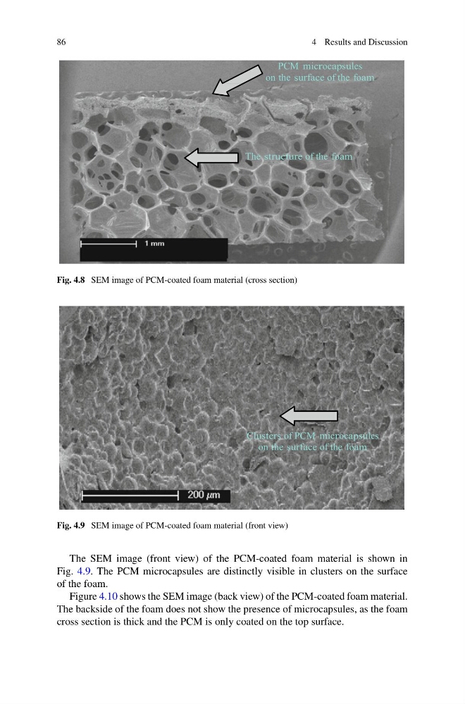
Fig. 4.8 SEM image of PCM-coated foam material (cross section)
Clusters of PCM microcapsules
on the surface of the foam
Fig. 4.9 SEM image of PCM-coated foam material (front view)
The SEM image (front view) of the PCM-coated foam material is shown in
Fig. 4.9. The PCM microcapsules are distinctly visible in clusters on the surface
of the foam.
Figure 4.10 shows the SEM image (back view) of the PCM-coated foam material.
The backside of the foam does not show the presence of microcapsules, as the foam
cross section is thick and the PCM is only coated on the top surface.
4 Results and Discussion
PCM microcapsules
on the surface of the foam
The structure of the foam
Fig. 4.8 SEM image of PCM-coated foam material (cross section)
Clusters of PCM microcapsules
on the surface of the foam
Fig. 4.9 SEM image of PCM-coated foam material (front view)
The SEM image (front view) of the PCM-coated foam material is shown in
Fig. 4.9. The PCM microcapsules are distinctly visible in clusters on the surface
of the foam.
Figure 4.10 shows the SEM image (back view) of the PCM-coated foam material.
The backside of the foam does not show the presence of microcapsules, as the foam
cross section is thick and the PCM is only coated on the top surface.