

4.1 SEM Images of the Samples
85
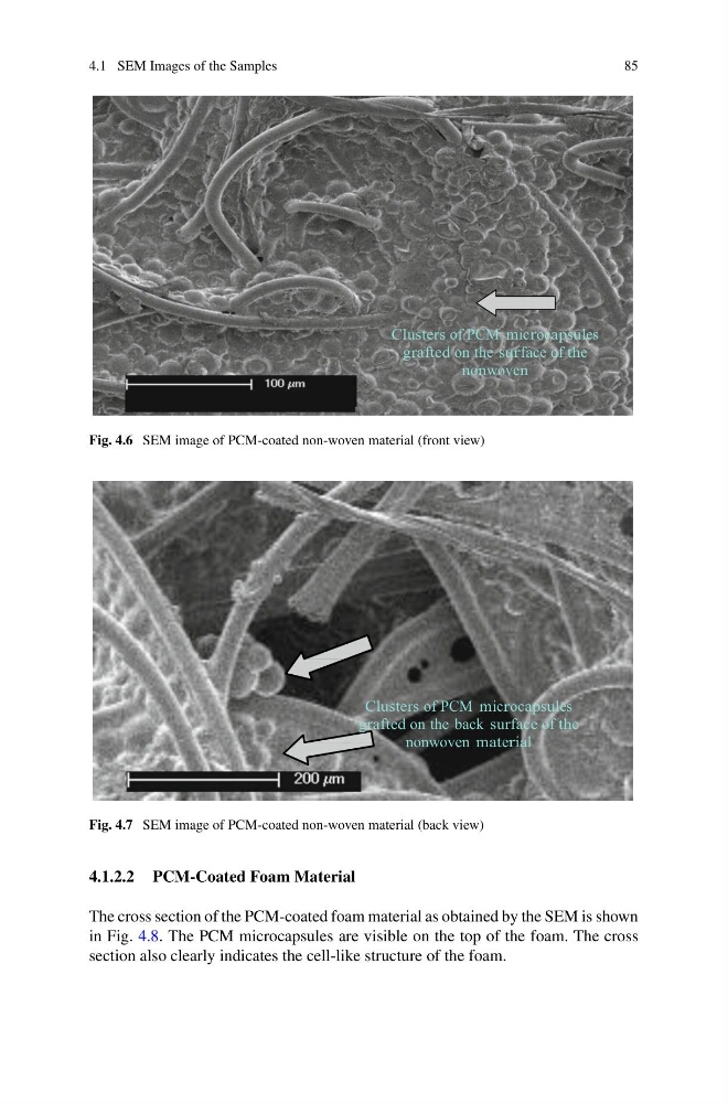
Clusters of PCM microcapsules
grafted on the surface of the
nonwoven
Fig. 4.6 SEM image of PCM-coated non-woven material (front view)
Clusters of PCM microcapsules
grafted on the back surface of the
nonwoven material
Fig. 4.7 SEM image of PCM-coated non-woven material (back view)
4.1.2.2 PCM-Coated Foam Material
The cross section of the PCM-coated foam material as obtained by the SEM is shown
in Fig. 4.8. The PCM microcapsules are visible on the top of the foam. The cross
section also clearly indicates the cell-like structure of the foam.
85
Clusters of PCM microcapsules
grafted on the surface of the
nonwoven
Fig. 4.6 SEM image of PCM-coated non-woven material (front view)
Clusters of PCM microcapsules
grafted on the back surface of the
nonwoven material
Fig. 4.7 SEM image of PCM-coated non-woven material (back view)
4.1.2.2 PCM-Coated Foam Material
The cross section of the PCM-coated foam material as obtained by the SEM is shown
in Fig. 4.8. The PCM microcapsules are visible on the top of the foam. The cross
section also clearly indicates the cell-like structure of the foam.