

78
5 Cells in Motion
There are different ways to crawl. The leading edge is often an almost twodimensional protrusion of the actin mesh, a “thin-sheet foot”, translated into biologists’ Latin as lamellipodium. It may inch ahead by tentatively protruding filopodia
– another foot in the term, though they are not feet at all but aids to explore the way
ahead: they can be either withdrawn or strengthened by actin filaments to pull the
leading edge ahead (Fig. 5.15). Nascent focal adhesions are formed at the lamellipodium and mature as the bulk of the cell advances. The actin network depolymerizes at the back end, and actin monomers are transported to the front to polymerize
there. This is a slow process limiting the speed to about the cell length per minute.
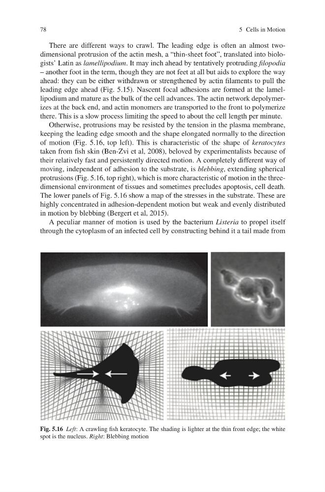
Otherwise, protrusions may be resisted by the tension in the plasma membrane,
keeping the leading edge smooth and the shape elongated normally to the direction
of motion (Fig. 5.16, top left). This is characteristic of the shape of keratocytes
taken from fish skin (Ben-Zvi et al, 2008), beloved by experimentalists because of
their relatively fast and persistently directed motion. A completely different way of
moving, independent of adhesion to the substrate, is blebbing, extending spherical
protrusions (Fig. 5.16, top right), which is more characteristic of motion in the threedimensional environment of tissues and sometimes precludes apoptosis, cell death.
The lower panels of Fig. 5.16 show a map of the stresses in the substrate. These are
highly concentrated in adhesion-dependent motion but weak and evenly distributed
in motion by blebbing (Bergert et al, 2015).
A peculiar manner of motion is used by the bacterium Listeria to propel itself
through the cytoplasm of an infected cell by constructing behind it a tail made from
Fig. 5.16 Left: A crawling fish keratocyte. The shading is lighter at the thin front edge; the white
spot is the nucleus. Right: Blebbing motion
5 Cells in Motion
There are different ways to crawl. The leading edge is often an almost twodimensional protrusion of the actin mesh, a “thin-sheet foot”, translated into biologists’ Latin as lamellipodium. It may inch ahead by tentatively protruding filopodia
– another foot in the term, though they are not feet at all but aids to explore the way
ahead: they can be either withdrawn or strengthened by actin filaments to pull the
leading edge ahead (Fig. 5.15). Nascent focal adhesions are formed at the lamellipodium and mature as the bulk of the cell advances. The actin network depolymerizes at the back end, and actin monomers are transported to the front to polymerize
there. This is a slow process limiting the speed to about the cell length per minute.
Otherwise, protrusions may be resisted by the tension in the plasma membrane,
keeping the leading edge smooth and the shape elongated normally to the direction
of motion (Fig. 5.16, top left). This is characteristic of the shape of keratocytes
taken from fish skin (Ben-Zvi et al, 2008), beloved by experimentalists because of
their relatively fast and persistently directed motion. A completely different way of
moving, independent of adhesion to the substrate, is blebbing, extending spherical
protrusions (Fig. 5.16, top right), which is more characteristic of motion in the threedimensional environment of tissues and sometimes precludes apoptosis, cell death.
The lower panels of Fig. 5.16 show a map of the stresses in the substrate. These are
highly concentrated in adhesion-dependent motion but weak and evenly distributed
in motion by blebbing (Bergert et al, 2015).
A peculiar manner of motion is used by the bacterium Listeria to propel itself
through the cytoplasm of an infected cell by constructing behind it a tail made from
Fig. 5.16 Left: A crawling fish keratocyte. The shading is lighter at the thin front edge; the white
spot is the nucleus. Right: Blebbing motion