

112
7 Communication
Ben-Jacob et al (1994) modified the model discussed above by adding chemotaxis in
the field of a signaling chemical, secreted or consumed by bacteria in a way that depended on their level of nutrition. This provided another interaction mechanism that
changed the shape of the colony with other parameters kept fixed, but one could find
a similar form among the patterns in Fig. 7.4. Designing various models of “active
media” became popular among theorists. Some of them produce dazzling patterns,
beating anything displayed by abstract expressionists. The review (Marchetti et al,
2013) coauthored by seven prominent physicists from three continents included 280
references and accumulated over a thousand citing papers over a period of six years,
of which only about 5% are directly related to biology.
Biologists are more concerned with biofilms where the embedding matrix inhibits motion but chemical interactions are pronounced to a degree approaching
multicellular organisms. The integrating mechanism is quorum sensing (Shapiro,
1998; Waters and Bassler, 2005). Bacteria detect signaling molecules and change
their gene expression, and hence also their behavior, when a certain threshold is
passed (Fig. 7.5). Biofilms usually include different microbe species, and bacteria
are able to differentiate between signals sent by their kin or non-kin. Cooperation
between bacteria, either genomically related or not, involves the metabolic costs of
signaling, which are rewarded by the benefits of division of labor that leads to more
efficient proliferation, and of collective defence against invaders or antibiotics. Interaction between bacterial species may even go as far as exchange of DNA, the
bacterial analogue of sex.
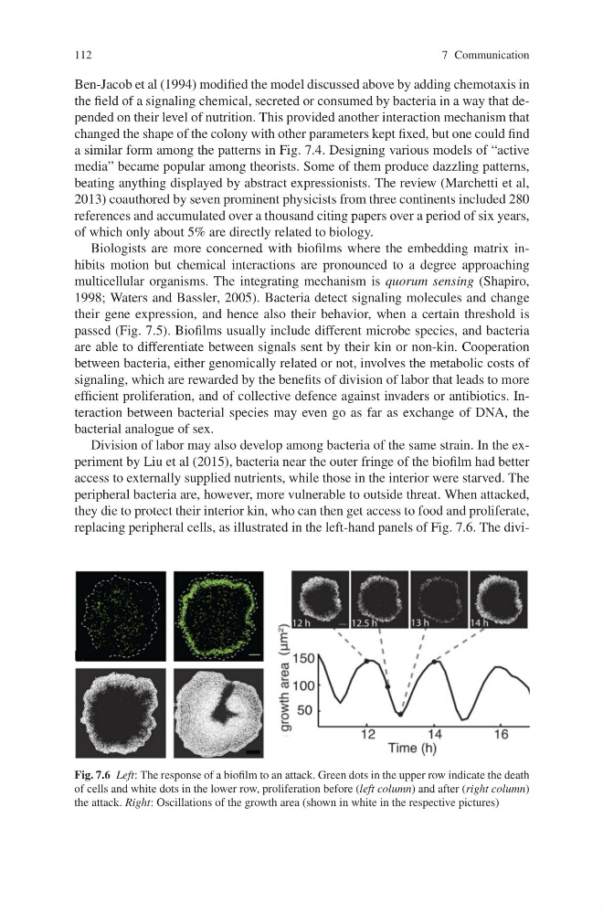
Division of labor may also develop among bacteria of the same strain. In the experiment by Liu et al (2015), bacteria near the outer fringe of the biofilm had better
access to externally supplied nutrients, while those in the interior were starved. The
peripheral bacteria are, however, more vulnerable to outside threat. When attacked,
they die to protect their interior kin, who can then get access to food and proliferate,
replacing peripheral cells, as illustrated in the left-hand panels of Fig. 7.6. The diviFig. 7.6 Left: The response of a biofilm to an attack. Green dots in the upper row indicate the death
of cells and white dots in the lower row, proliferation before (left column) and after (right column)
the attack. Right: Oscillations of the growth area (shown in white in the respective pictures)
7 Communication
Ben-Jacob et al (1994) modified the model discussed above by adding chemotaxis in
the field of a signaling chemical, secreted or consumed by bacteria in a way that depended on their level of nutrition. This provided another interaction mechanism that
changed the shape of the colony with other parameters kept fixed, but one could find
a similar form among the patterns in Fig. 7.4. Designing various models of “active
media” became popular among theorists. Some of them produce dazzling patterns,
beating anything displayed by abstract expressionists. The review (Marchetti et al,
2013) coauthored by seven prominent physicists from three continents included 280
references and accumulated over a thousand citing papers over a period of six years,
of which only about 5% are directly related to biology.
Biologists are more concerned with biofilms where the embedding matrix inhibits motion but chemical interactions are pronounced to a degree approaching
multicellular organisms. The integrating mechanism is quorum sensing (Shapiro,
1998; Waters and Bassler, 2005). Bacteria detect signaling molecules and change
their gene expression, and hence also their behavior, when a certain threshold is
passed (Fig. 7.5). Biofilms usually include different microbe species, and bacteria
are able to differentiate between signals sent by their kin or non-kin. Cooperation
between bacteria, either genomically related or not, involves the metabolic costs of
signaling, which are rewarded by the benefits of division of labor that leads to more
efficient proliferation, and of collective defence against invaders or antibiotics. Interaction between bacterial species may even go as far as exchange of DNA, the
bacterial analogue of sex.
Division of labor may also develop among bacteria of the same strain. In the experiment by Liu et al (2015), bacteria near the outer fringe of the biofilm had better
access to externally supplied nutrients, while those in the interior were starved. The
peripheral bacteria are, however, more vulnerable to outside threat. When attacked,
they die to protect their interior kin, who can then get access to food and proliferate,
replacing peripheral cells, as illustrated in the left-hand panels of Fig. 7.6. The diviFig. 7.6 Left: The response of a biofilm to an attack. Green dots in the upper row indicate the death
of cells and white dots in the lower row, proliferation before (left column) and after (right column)
the attack. Right: Oscillations of the growth area (shown in white in the respective pictures)