

96
6 Cells United
the volume changes, and this costs energy. The perimeter increases at a fixed area
if the shape of the cell becomes irregular. Vertices move to decrease the overall
energy, and the derivative of the energy with respect to the position of the vortex is
interpreted as the driving force. Any external force, if present, can be added here,
and it may counteract the tendency of the layer to relax to a regular pattern.
This is, of course, a very rough way to overcome the complexities of restructuring
the cytoskeleton of cells that has to accompany their motion, but it produces realistically distorted cell shapes when combined with cell division and intercalation. The
latter process, shown schematically in the left-hand panel of Fig. 6.10, may naturally
lead to elongation and narrowing of the tissue when a force is applied in a certain direction, or when the cells grow or multiply while laterally confined, as demonstrated
by the transition between the configurations in the central and right-hand panels of
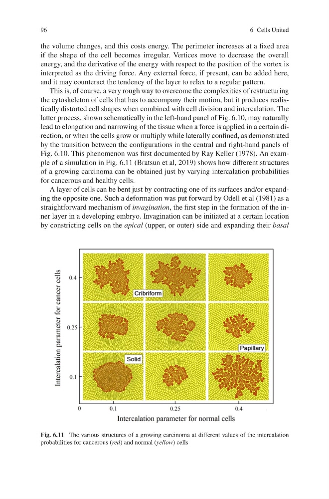
Fig. 6.10. This phenomenon was first documented by Ray Keller (1978). An example of a simulation in Fig. 6.11 (Bratsun et al, 2019) shows how different structures
of a growing carcinoma can be obtained just by varying intercalation probabilities
for cancerous and healthy cells.
A layer of cells can be bent just by contracting one of its surfaces and/or expanding the opposite one. Such a deformation was put forward by Odell et al (1981) as a
straightforward mechanism of invagination, the first step in the formation of the inner layer in a developing embryo. Invagination can be initiated at a certain location
by constricting cells on the apical (upper, or outer) side and expanding their basal
Fig. 6.11 The various structures of a growing carcinoma at different values of the intercalation
probabilities for cancerous (red) and normal (yellow) cells
6 Cells United
the volume changes, and this costs energy. The perimeter increases at a fixed area
if the shape of the cell becomes irregular. Vertices move to decrease the overall
energy, and the derivative of the energy with respect to the position of the vortex is
interpreted as the driving force. Any external force, if present, can be added here,
and it may counteract the tendency of the layer to relax to a regular pattern.
This is, of course, a very rough way to overcome the complexities of restructuring
the cytoskeleton of cells that has to accompany their motion, but it produces realistically distorted cell shapes when combined with cell division and intercalation. The
latter process, shown schematically in the left-hand panel of Fig. 6.10, may naturally
lead to elongation and narrowing of the tissue when a force is applied in a certain direction, or when the cells grow or multiply while laterally confined, as demonstrated
by the transition between the configurations in the central and right-hand panels of
Fig. 6.10. This phenomenon was first documented by Ray Keller (1978). An example of a simulation in Fig. 6.11 (Bratsun et al, 2019) shows how different structures
of a growing carcinoma can be obtained just by varying intercalation probabilities
for cancerous and healthy cells.
A layer of cells can be bent just by contracting one of its surfaces and/or expanding the opposite one. Such a deformation was put forward by Odell et al (1981) as a
straightforward mechanism of invagination, the first step in the formation of the inner layer in a developing embryo. Invagination can be initiated at a certain location
by constricting cells on the apical (upper, or outer) side and expanding their basal
Fig. 6.11 The various structures of a growing carcinoma at different values of the intercalation
probabilities for cancerous (red) and normal (yellow) cells