

We also adopted a similar theoretical strategy for MurD and uridine
monophosphate kinase as druggable Mtb targets for curing TB disease [35, 36].
Recently, another key target in this family Mtb phospho-MurNAc-pentapeptide
translocase (also termed as MurX) involved in the PG biosynthesis was identified
and a sansanmycin uridyl peptide natural product analogue was discovered to be
selective and potent inhibitor by in vitro and intracellular anti-mycobacterial
activity assay [39]. The biggest challenge in the development of inhibitors targeting
Mur ligase enzymes will be the chemical structure optimization of the compounds
to enable the passage through the cell wall of Mtb and its stability in the cytoplasm.
Thus, in vivo and clinically successful compounds are yet to be discovered based on
the MurC ligases [40, 41].
Next key targets in almost all Gram-positive and Gram-negative bacterial species
are involved in the PG biosynthesis. Mtb PG is composed of a repeating disaccharide sugar units consisting of N-glycolylmuramic acid (MurNGlyc) linked to
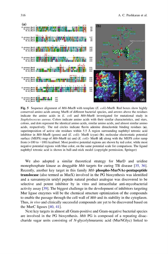
Fig. 5 Sequence alignment of Mtb-MurB with template (E. coli)-MurB. Red boxes show highly
conserved amino acids among MurB of different bacterial species, and arrows above the residues
indicate the amino acids in E. coli and Mtb-MurB investigated for mutational study in
Staphylococcus aureus. Colors indicate amino acids with their similar characteristics, and stars,
colons, and dots represent the identical amino acids, similar amino acids, and almost similar amino
acids, respectively. The red circles indicate flavin adenine dinucleotide binding residues (a),
superimposition of active site residues within 5.5 Å region surrounding naphthyl tetronic acid
inhibitor in Mtb-MurB (green) and (E. coli)- MurB (cyan) (b); molecular electrostatic potential
surface (MEPS) map of Mtb-MurB (c) and (E. coli)- MurB (d) along with the MEPS color ramp
from (+100 to −100) kcal/mol. Most positive potential regions are shown by red color, while most
negative potential regions with blue color, on the same potential scale for comparison. The ligand
naphthyl tetronic acid is shown in ball-and-stick model (copyright permission, Springer)
316
A. C. Pushkaran et al.
monophosphate kinase as druggable Mtb targets for curing TB disease [35, 36].
Recently, another key target in this family Mtb phospho-MurNAc-pentapeptide
translocase (also termed as MurX) involved in the PG biosynthesis was identified
and a sansanmycin uridyl peptide natural product analogue was discovered to be
selective and potent inhibitor by in vitro and intracellular anti-mycobacterial
activity assay [39]. The biggest challenge in the development of inhibitors targeting
Mur ligase enzymes will be the chemical structure optimization of the compounds
to enable the passage through the cell wall of Mtb and its stability in the cytoplasm.
Thus, in vivo and clinically successful compounds are yet to be discovered based on
the MurC ligases [40, 41].
Next key targets in almost all Gram-positive and Gram-negative bacterial species
are involved in the PG biosynthesis. Mtb PG is composed of a repeating disaccharide sugar units consisting of N-glycolylmuramic acid (MurNGlyc) linked to
Fig. 5 Sequence alignment of Mtb-MurB with template (E. coli)-MurB. Red boxes show highly
conserved amino acids among MurB of different bacterial species, and arrows above the residues
indicate the amino acids in E. coli and Mtb-MurB investigated for mutational study in
Staphylococcus aureus. Colors indicate amino acids with their similar characteristics, and stars,
colons, and dots represent the identical amino acids, similar amino acids, and almost similar amino
acids, respectively. The red circles indicate flavin adenine dinucleotide binding residues (a),
superimposition of active site residues within 5.5 Å region surrounding naphthyl tetronic acid
inhibitor in Mtb-MurB (green) and (E. coli)- MurB (cyan) (b); molecular electrostatic potential
surface (MEPS) map of Mtb-MurB (c) and (E. coli)- MurB (d) along with the MEPS color ramp
from (+100 to −100) kcal/mol. Most positive potential regions are shown by red color, while most
negative potential regions with blue color, on the same potential scale for comparison. The ligand
naphthyl tetronic acid is shown in ball-and-stick model (copyright permission, Springer)
316
A. C. Pushkaran et al.