

datasets [22, 23]. Further, the MM-PBSA method has been extensively used to
understand the interaction of various substrates with the targets that are relevant in
the treatment of various neurodegenerative diseases. A detailed account of this can
be found in the reference [22].
4.1 Molecular Docking
Molecular docking is the most simplistic method available for computing the
protein–ligand binding affinities and for finding the most stable binding mode
(pose) for a ligand within the binding site of the protein. The scoring functions are
used to decide on the binders from non-binders and their least energy binding mode
and pose which can be knowledge-based, empirical and force-field-based [24–26].
In the force-field-based scoring function, the free energy of binding dictates the
drug potency. The interaction energies are calculated as a sum of polar and
non-polar interactions such as van der Waals and electrostatic. The change in
intramolecular energies of the ligand due to conformational change is also added to
the total energies just to make sure ligand conformations with unusually high
energies are avoided in the search. The entropic contributions due to conformational
degree of freedom are included in a simple mean; i.e. each flexible bond is associated with 0.3 kcal/mol.
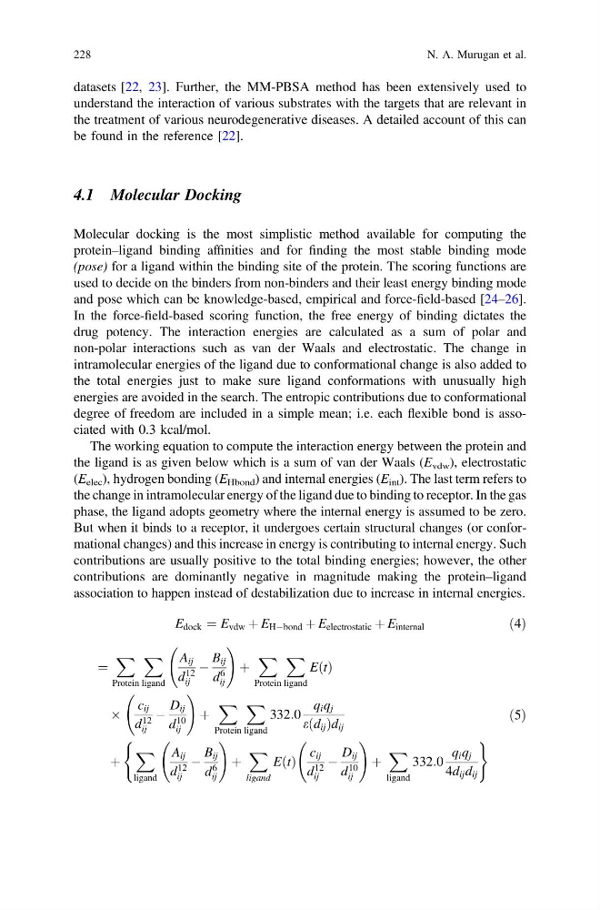
The working equation to compute the interaction energy between the protein and
the ligand is as given below which is a sum of van der Waals (E vdw ), electrostatic
(E elec ), hydrogen bonding (E Hbond ) and internal energies (E int ). The last term refers to
the change in intramolecular energy of the ligand due to binding to receptor. In the gas
phase, the ligand adopts geometry where the internal energy is assumed to be zero.
But when it binds to a receptor, it undergoes certain structural changes (or conformational changes) and this increase in energy is contributing to internal energy. Such
contributions are usually positive to the total binding energies; however, the other
contributions are dominantly negative in magnitude making the protein–ligand
association to happen instead of destabilization due to increase in internal energies.
E dock ¼ E vdw þ E HÀbond þ E electrostatic þ E internal
ð4Þ
¼
X
Protein
X
ligand
A ij
d 12
ij
À
B ij
d
6
ij
!
þ
X
Protein
X
ligand
EðtÞ
Â
c ij
d 12
ij
À
D ij
d
10
ij
!
þ
X
Protein
X
ligand
332:0
q i q j
eðd ij Þd ij
þ
X
ligand
A ij
d 12
ij
À
B ij
d
6
ij
!
þ
X
ligand
EðtÞ
c ij
d 12
ij
À
D ij
d
10
ij
!
þ
X
ligand
332:0
q i q j
4d ij d ij
(
)
ð5Þ
228
N. A. Murugan et al.
understand the interaction of various substrates with the targets that are relevant in
the treatment of various neurodegenerative diseases. A detailed account of this can
be found in the reference [22].
4.1 Molecular Docking
Molecular docking is the most simplistic method available for computing the
protein–ligand binding affinities and for finding the most stable binding mode
(pose) for a ligand within the binding site of the protein. The scoring functions are
used to decide on the binders from non-binders and their least energy binding mode
and pose which can be knowledge-based, empirical and force-field-based [24–26].
In the force-field-based scoring function, the free energy of binding dictates the
drug potency. The interaction energies are calculated as a sum of polar and
non-polar interactions such as van der Waals and electrostatic. The change in
intramolecular energies of the ligand due to conformational change is also added to
the total energies just to make sure ligand conformations with unusually high
energies are avoided in the search. The entropic contributions due to conformational
degree of freedom are included in a simple mean; i.e. each flexible bond is associated with 0.3 kcal/mol.
The working equation to compute the interaction energy between the protein and
the ligand is as given below which is a sum of van der Waals (E vdw ), electrostatic
(E elec ), hydrogen bonding (E Hbond ) and internal energies (E int ). The last term refers to
the change in intramolecular energy of the ligand due to binding to receptor. In the gas
phase, the ligand adopts geometry where the internal energy is assumed to be zero.
But when it binds to a receptor, it undergoes certain structural changes (or conformational changes) and this increase in energy is contributing to internal energy. Such
contributions are usually positive to the total binding energies; however, the other
contributions are dominantly negative in magnitude making the protein–ligand
association to happen instead of destabilization due to increase in internal energies.
E dock ¼ E vdw þ E HÀbond þ E electrostatic þ E internal
ð4Þ
¼
X
Protein
X
ligand
A ij
d 12
ij
À
B ij
d
6
ij
!
þ
X
Protein
X
ligand
EðtÞ
Â
c ij
d 12
ij
À
D ij
d
10
ij
!
þ
X
Protein
X
ligand
332:0
q i q j
eðd ij Þd ij
þ
X
ligand
A ij
d 12
ij
À
B ij
d
6
ij
!
þ
X
ligand
EðtÞ
c ij
d 12
ij
À
D ij
d
10
ij
!
þ
X
ligand
332:0
q i q j
4d ij d ij
(
)
ð5Þ
228
N. A. Murugan et al.