

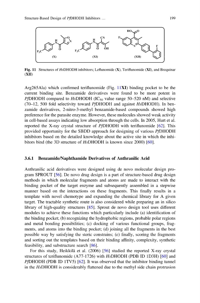
Arg265Ala) which confirmed teriflunomide (Fig. 11XI) binding pocket to be the
current binding site. Benzamide derivatives were found to be more potent in
PfDHODH compared to HsDHODH (IC 50 value range 50–520 nM) and selective
(70–12, 500 fold selectivity toward PfDHODH and against HsDHODH). In benzamide derivatives, 2-nitro-3-methyl benzamide-based compounds showed high
preference for the parasite enzyme. However, these molecules showed weak activity
in cell-based assays indicating low absorption through the cells. In 2005, Hurt et al.
reported the X-ray crystal structure of PfDHODH with teriflunomide [62]. This
provided opportunity for the SBDD approach for designing of various PfDHODH
inhibitors based on the detailed knowledge about the active site in which the inhibitors bind (the 3D structure of HsDHODH is known since 2000) [60].
3.6.1 Benzamide/Naphthamide Derivatives of Anthranilic Acid
Anthranilic acid derivatives were designed using de novo molecular design program SPROUT [56]. De novo drug design is a part of structure-based drug design
methods in which molecular fragments and atoms are made to interact with the
binding pocket of the target enzyme and subsequently assembled in a stepwise
manner based on the interactions on these fragments. This finally results in a
template with novel chemotype and expanding the chemical library for A given
target. The tractable synthetic route is also considered while preparing an in silico
library of high-quality structures [85]. Sprout de novo design tool uses different
modules to achieve these functions which particularly include (a) identification of
the binding pocket; (b) recognizing the hydrophobic regions, probable polar regions
and metal bonding possibilities; (c) docking of various functional groups, fragments, and atoms into the binding pocket; (d) joining all the fragments in the best
possible way by satisfying the steric constrains; (e) finally, scoring the fragments
and sorting out the templates based on their binding affinity, complexity, synthetic
feasibility, and substructure search [86].
For this study, Heikkilä et al. (2006) [56] studied the reported X-ray crystal
structures of teriflunomide (A77-1726) with HsDHODH (PDB ID 1D3H) [60] and
PfDHODH (PDB ID 1TV5) [62]. It was observed that the inhibitor binding tunnel
in the HsDHODH is considerably flattened due to the methyl side chain protrusion
Fig. 11 Structures of HsDHODH inhibitors; Leflunomide (X), Teriflunomide (XI), and Brequinar
(XII)
Structure-Based Design of PfDHODH Inhibitors …
199
current binding site. Benzamide derivatives were found to be more potent in
PfDHODH compared to HsDHODH (IC 50 value range 50–520 nM) and selective
(70–12, 500 fold selectivity toward PfDHODH and against HsDHODH). In benzamide derivatives, 2-nitro-3-methyl benzamide-based compounds showed high
preference for the parasite enzyme. However, these molecules showed weak activity
in cell-based assays indicating low absorption through the cells. In 2005, Hurt et al.
reported the X-ray crystal structure of PfDHODH with teriflunomide [62]. This
provided opportunity for the SBDD approach for designing of various PfDHODH
inhibitors based on the detailed knowledge about the active site in which the inhibitors bind (the 3D structure of HsDHODH is known since 2000) [60].
3.6.1 Benzamide/Naphthamide Derivatives of Anthranilic Acid
Anthranilic acid derivatives were designed using de novo molecular design program SPROUT [56]. De novo drug design is a part of structure-based drug design
methods in which molecular fragments and atoms are made to interact with the
binding pocket of the target enzyme and subsequently assembled in a stepwise
manner based on the interactions on these fragments. This finally results in a
template with novel chemotype and expanding the chemical library for A given
target. The tractable synthetic route is also considered while preparing an in silico
library of high-quality structures [85]. Sprout de novo design tool uses different
modules to achieve these functions which particularly include (a) identification of
the binding pocket; (b) recognizing the hydrophobic regions, probable polar regions
and metal bonding possibilities; (c) docking of various functional groups, fragments, and atoms into the binding pocket; (d) joining all the fragments in the best
possible way by satisfying the steric constrains; (e) finally, scoring the fragments
and sorting out the templates based on their binding affinity, complexity, synthetic
feasibility, and substructure search [86].
For this study, Heikkilä et al. (2006) [56] studied the reported X-ray crystal
structures of teriflunomide (A77-1726) with HsDHODH (PDB ID 1D3H) [60] and
PfDHODH (PDB ID 1TV5) [62]. It was observed that the inhibitor binding tunnel
in the HsDHODH is considerably flattened due to the methyl side chain protrusion
Fig. 11 Structures of HsDHODH inhibitors; Leflunomide (X), Teriflunomide (XI), and Brequinar
(XII)
Structure-Based Design of PfDHODH Inhibitors …
199