

favorable). Again, when looking at the geometric quality criterion for two structures, 1FQX only (chain A: 51% and chain B: 52% residues) does not have any
outlier, while rest (chain A: 47% and chain B: 40%) have at least one outlier. The
geometric quality for 4ZIP seems better as in this case 96 and 90% residues (chains
A and B, respectively) do not have any geometric outlier. Further considering fit
quality of the model to electron density, 1FQX has certain residues which has at
least two outliers and a significant percentage of residues with at least one outlier,
while in case of 4ZIP, there are no residues which have two outliers and only a
small fraction of residues with only single outlier. Considering all above points
among 1FQX and 4ZIP, 4ZIP should be preferable over 1FQX as receptor structure
for any docking study.
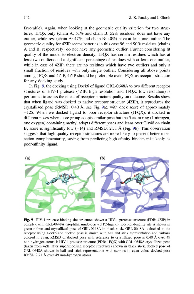
In Fig. 9, the docking using Dock6 of ligand GRL-0648A to two different receptor
structures of HIV-1 protease (4ZIP: high resolution and 1FQX: low resolution) is
performed to assess the effect of receptor structure quality on outcome. Results show
that when ligand was docked to native receptor structure (4ZIP), it reproduces the
crystallized pose (RMSD: 0.40 Å, see Fig. 9a), with dock score of approximately
−125. When we docked ligand to poor receptor structure (1FQX), it docked in
different poses where core group adopts similar pose but the 5-atom ring (1 nitrogen,
one oxygen) containing methyl adopts different poses and leans over Gly48 on chain
B, score is significantly low (−14) and RMSD: 2.71 Å (Fig. 9b). This observation
suggests that high-quality receptor structures are more likely to present better interaction complementarity, saving from predicting high-affinity binders mistakenly as
poor-affinity ligand.
Fig. 9 HIV-1 protease-binding site structures shown a HIV-1 protease structure (PDB: 4ZIP) in
complex with GRL-0648A (isophthalamide-derived P2-ligand), receptor-binding site is shown in
green ribbon and crystallized pose of GRL-0648A in black stick. GRL-0648A is docked to the
receptor using Dock6 and docked pose is shown with ball and stick representation and carbons
colored in cyan, RMSD of docked pose with reference to crystallized pose is 0.40 Å over 49
non-hydrogen atoms. b HIV-1 protease structure (PDB: 1FQX) with GRL-0648A crystallized pose
(taken from 4ZIP after superimposing receptor structures) shown in black stick, docked pose of
GRL-0648A shown in ball and stick representation with carbons in cyan color, docked pose
RMSD 2.71 Å over 49 non-hydrogen atoms
142
S. K. Panday and I. Ghosh
outlier, while rest (chain A: 47% and chain B: 40%) have at least one outlier. The
geometric quality for 4ZIP seems better as in this case 96 and 90% residues (chains
A and B, respectively) do not have any geometric outlier. Further considering fit
quality of the model to electron density, 1FQX has certain residues which has at
least two outliers and a significant percentage of residues with at least one outlier,
while in case of 4ZIP, there are no residues which have two outliers and only a
small fraction of residues with only single outlier. Considering all above points
among 1FQX and 4ZIP, 4ZIP should be preferable over 1FQX as receptor structure
for any docking study.
In Fig. 9, the docking using Dock6 of ligand GRL-0648A to two different receptor
structures of HIV-1 protease (4ZIP: high resolution and 1FQX: low resolution) is
performed to assess the effect of receptor structure quality on outcome. Results show
that when ligand was docked to native receptor structure (4ZIP), it reproduces the
crystallized pose (RMSD: 0.40 Å, see Fig. 9a), with dock score of approximately
−125. When we docked ligand to poor receptor structure (1FQX), it docked in
different poses where core group adopts similar pose but the 5-atom ring (1 nitrogen,
one oxygen) containing methyl adopts different poses and leans over Gly48 on chain
B, score is significantly low (−14) and RMSD: 2.71 Å (Fig. 9b). This observation
suggests that high-quality receptor structures are more likely to present better interaction complementarity, saving from predicting high-affinity binders mistakenly as
poor-affinity ligand.
Fig. 9 HIV-1 protease-binding site structures shown a HIV-1 protease structure (PDB: 4ZIP) in
complex with GRL-0648A (isophthalamide-derived P2-ligand), receptor-binding site is shown in
green ribbon and crystallized pose of GRL-0648A in black stick. GRL-0648A is docked to the
receptor using Dock6 and docked pose is shown with ball and stick representation and carbons
colored in cyan, RMSD of docked pose with reference to crystallized pose is 0.40 Å over 49
non-hydrogen atoms. b HIV-1 protease structure (PDB: 1FQX) with GRL-0648A crystallized pose
(taken from 4ZIP after superimposing receptor structures) shown in black stick, docked pose of
GRL-0648A shown in ball and stick representation with carbons in cyan color, docked pose
RMSD 2.71 Å over 49 non-hydrogen atoms
142
S. K. Panday and I. Ghosh