

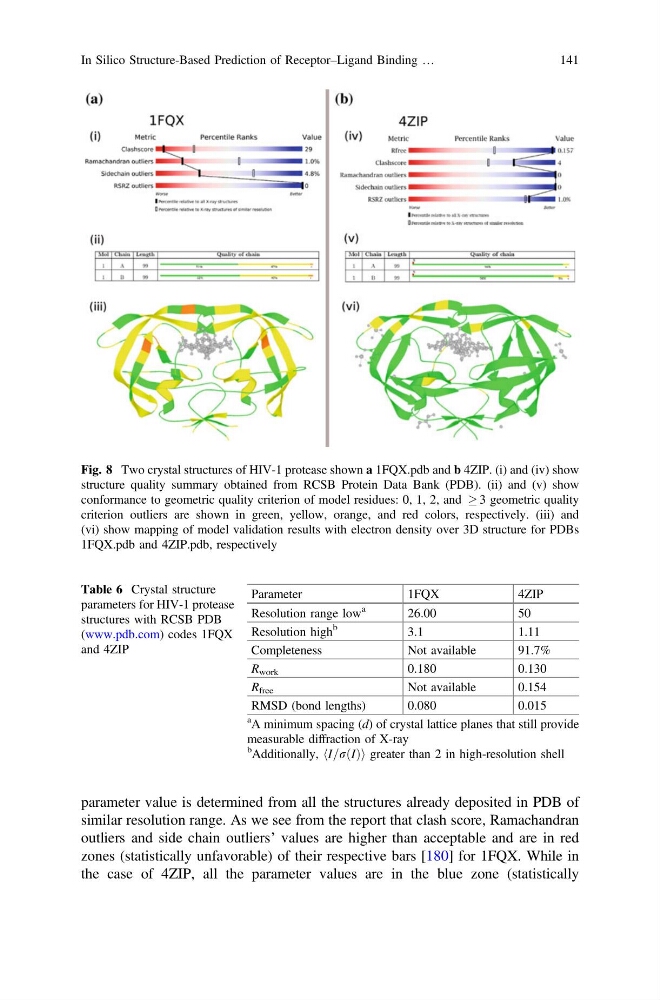
parameter value is determined from all the structures already deposited in PDB of
similar resolution range. As we see from the report that clash score, Ramachandran
outliers and side chain outliers’ values are higher than acceptable and are in red
zones (statistically unfavorable) of their respective bars [180] for 1FQX. While in
the case of 4ZIP, all the parameter values are in the blue zone (statistically
Fig. 8 Two crystal structures of HIV-1 protease shown a 1FQX.pdb and b 4ZIP. (i) and (iv) show
structure quality summary obtained from RCSB Protein Data Bank (PDB). (ii) and (v) show
conformance to geometric quality criterion of model residues: 0, 1, 2, and ! 3 geometric quality
criterion outliers are shown in green, yellow, orange, and red colors, respectively. (iii) and
(vi) show mapping of model validation results with electron density over 3D structure for PDBs
1FQX.pdb and 4ZIP.pdb, respectively
Table 6 Crystal structure
parameters for HIV-1 protease
structures with RCSB PDB
(www.pdb.com) codes 1FQX
and 4ZIP
Parameter
1FQX
4ZIP
Resolution range low
a
26.00
50
Resolution high
b
3.1
1.11
Completeness
Not available
91.7%
R work
0.180
0.130
R free
Not available
0.154
RMSD (bond lengths)
0.080
0.015
a A minimum spacing (d) of crystal lattice planes that still provide
measurable diffraction of X-ray
b
Additionally, hI=r I
ð Þi greater than 2 in high-resolution shell
In Silico Structure-Based Prediction of Receptor–Ligand Binding …
141
similar resolution range. As we see from the report that clash score, Ramachandran
outliers and side chain outliers’ values are higher than acceptable and are in red
zones (statistically unfavorable) of their respective bars [180] for 1FQX. While in
the case of 4ZIP, all the parameter values are in the blue zone (statistically
Fig. 8 Two crystal structures of HIV-1 protease shown a 1FQX.pdb and b 4ZIP. (i) and (iv) show
structure quality summary obtained from RCSB Protein Data Bank (PDB). (ii) and (v) show
conformance to geometric quality criterion of model residues: 0, 1, 2, and ! 3 geometric quality
criterion outliers are shown in green, yellow, orange, and red colors, respectively. (iii) and
(vi) show mapping of model validation results with electron density over 3D structure for PDBs
1FQX.pdb and 4ZIP.pdb, respectively
Table 6 Crystal structure
parameters for HIV-1 protease
structures with RCSB PDB
(www.pdb.com) codes 1FQX
and 4ZIP
Parameter
1FQX
4ZIP
Resolution range low
a
26.00
50
Resolution high
b
3.1
1.11
Completeness
Not available
91.7%
R work
0.180
0.130
R free
Not available
0.154
RMSD (bond lengths)
0.080
0.015
a A minimum spacing (d) of crystal lattice planes that still provide
measurable diffraction of X-ray
b
Additionally, hI=r I
ð Þi greater than 2 in high-resolution shell
In Silico Structure-Based Prediction of Receptor–Ligand Binding …
141